
|
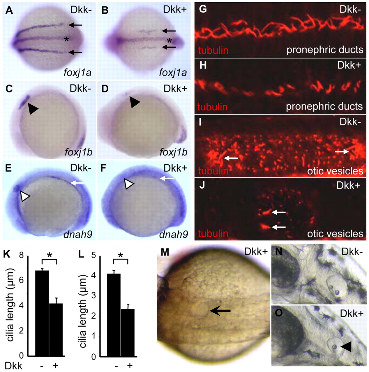
Fig. 6 Dkk1 induction impairs ciliogenesis in PD and OV. (A-D) Induction of Dkk1 downregulates foxj1 expression in PDs and OVs. foxj1a level in PDs was downregulated in transgenic Dkk1 embryos (B; 8/8) versus non-transgenic siblings (A). foxj1a expression in FP was not affected (B). foxj1b expression in the otic placode was downregulated in transgenic Dkk1 embryos (D; 8/8) versus non-transgenic siblings (C). Arrow indicates PDs, asterisk FP, and arrowhead the otic placode. (E,F) Induction of Dkk1 suppressed dnah9 expression in PDs and OVs (F; 11/11) compared with controls (E). White arrow represents PDs, and white arrowhead the otic placode. Shown are dorsal views (A,B) and lateral views (C-F) of 10-somite staged embryos with anterior to the left. (G-J) Induction of Dkk1 results in shorter and fewer cilia in PDs and OVs. Cilia were visualized by anti-tubulin antibody staining of 26-somite staged embryos. White arrow indicates tethering cilia in OVs. (K,L) Quantification of cilia length in PDs (K) and OVs (tethering cilia) (L). Data are represented as mean±s.d. Approximately 9-18 embryos were used for each group. *P<0.01. (M-O) Induction of Dkk1 results in kidney cysts and otolith malformation. Cystic distension of PD (arrow in M; 45/73) was observed at 24 hpf. One otolith (arrowhead in O; 89/95) was seen at 2 dpf. Tg(hsp:dkk1-GFP) embryos were heat activated at 60% epiboly.