
Image
|
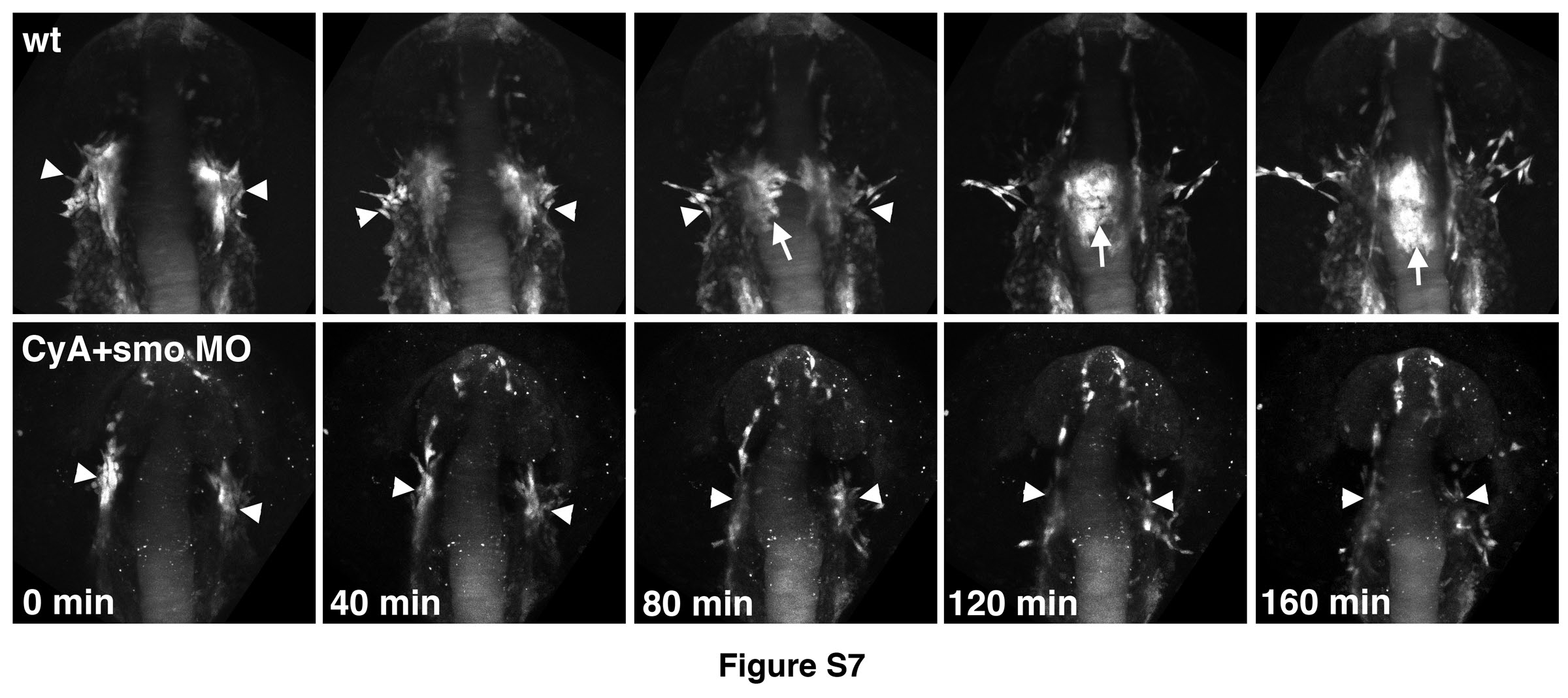
Figure Caption
Fig. S7
Time-lapse images of endocardial formation in etsrp:GFP control and CyAtreated smo morphant embryos.
Embryos were imaged starting from the 11-12-somite stage. Dorsal view of the cranial region, anterior is up. Etsrp:GFP-expression labels vascular endothelial, myeloid and endocardial lineages that originate primarily in the midbrain organizing center (MOC, arrowheads). Note that MOC formation is not significantly affected in CyA+smo MO embryos (0 min). Endocardial progenitors (arrow) migrate to the midline in wt embryos while this migration is largely absent in CyA+smo morphants.
Acknowledgments
This image is the copyrighted work of the attributed author or publisher, and
ZFIN has permission only to display this image to its users.
Additional permissions should be obtained from the applicable author or publisher of the image.
Reprinted from Developmental Biology, 361(2), Wong, K.S., Rehn, K., Palencia-Desai, S., Kohli, V., Hunter, W., Uhl, J.D., Rost, M.S., and Sumanas, S., Hedgehog signaling is required for differentiation of endocardial progenitors in zebrafish, 377-91, Copyright (2012) with permission from Elsevier. Full text @ Dev. Biol.