
|
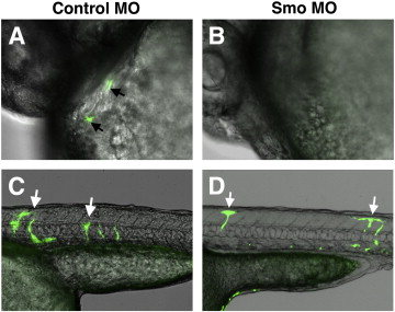
Fig. 11 Donor cells from smo MO-injected Tg(fli1a:EGFP) embryos contribute to vascular endothelial but not endocardial lineage. Cells were transplanted from smo MO or control MO-injected Tg(fli1a:EGFP) embryos into wt recipients. Overlap of bright field and green channels, anterior is to the left, embryos are at 32 hpf. (A,B) Endocardial cells from Tg(fli1a:EGFP) control donor embryos (arrows, A) contribute at a greater frequency to the endocardium of wt recipient embryos compared to the cells from smo MO donors (B). Note that while two fluorescent cells are shown in (A), the majority of recipient embryos that showed any contribution of Tg(fli1a:EGFP) cells to the endocardium, displayed only a single GFP-positive cell. (C,D) Transplanted cells from both control and smo morphants contribute to vascular endothelium (arrows).
Reprinted from Developmental Biology, 361(2), Wong, K.S., Rehn, K., Palencia-Desai, S., Kohli, V., Hunter, W., Uhl, J.D., Rost, M.S., and Sumanas, S., Hedgehog signaling is required for differentiation of endocardial progenitors in zebrafish, 377-91, Copyright (2012) with permission from Elsevier. Full text @ Dev. Biol.