
|
Fig. S2
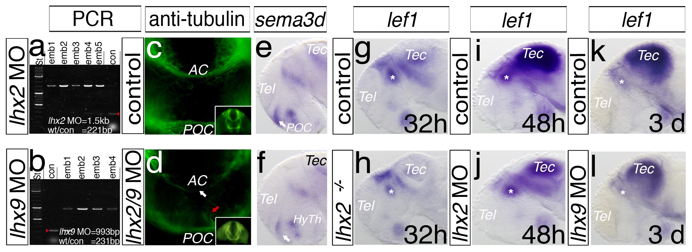
Efficient knock-down of lhx2 and lhx9 during forebrain development. To validate the efficiency of the lhx2 and lhx9 splice-site Morpholino-antisense oligomere approach, we isolated cDNA from injected and non-injected embryos at 48 hpf. A PCR approach, with primers flanking exon1 and exon2 of lhx2, demonstrates a suppression of the splicing event of intron1 (1.5 kb) in five individual embryos injected with lhx2 MO (emb1–5) compared to a control embryo (con, 221 bp) (a). A similar effect is demonstrated in injected lhx9 MO embryos 1–4 (emb1–4; b), which display a non-splicing event of intron1 (993 bp), compared to control embryos (con, 231 bp) (b). An antibody against acetylated tubulin shows midline crossing axons anterior (AC, anterior commissure) and posterior (POC, post-optic commissure) in the telencephalon (c). In lhx2/lhx9 double morphant embryos, both commissures do not cross the midline (arrow, d). Single in situ hybridizations of embryos at 48 hpf are displayed by a lateral view (e–l). Knock-down of Lhx2 and Lhx9 leads to a decrease of sema3d expression in postoptic commissure (POC, arrow; e, f). The morphant analysis of single knock-down, either lhx2 or lhx9, shows that lef1 expression is unaltered in the thalamus (h, j), compared to the control embryos (g, i, k). In the lhx2 mutant embryos, lef1 expression in the thalamus shows a weak alteration (l). HyTh, hypothalamus; MDO, mid-diencephalic-organizer; pTu, posterior tuberculum; RP, roof plate; Tec, tectum; Tel, telencephalon.