
|
Fig. 7
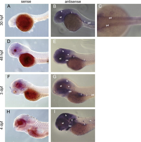
Whole mount in situ hybridization shows distinct expression of 20beta-HSD type 2 during zebrafish development. In 30 hpf embryos (B and C), expression is observed in brain, eye, ear, intestine, liver, and pronephric ducts. In 48 hpf (E) and 3 dpf larvae (G), signals are detected in all parts of brain, eye, intestine, liver, pronephric ducts, and branchial arches. 4 dpf larvae (I) show expression in brain, eye, ear, pharyngeal arches, intestine, liver, pronephric ducts, and neuromasts. Specific hybridization is indicated by blue color. Control hybridizations with sense probe are shown for all stages (30 hpf (A), 48 hpf (D), 3 dpf (F) and 4 dpf (H)). hpf, hours post fertilization; dpf, days post fertilization; b, brain; el, eye and lens; e, ear; l, liver; i, intestine; pd, pronephric ducts; pa, pharyngeal arches; ba, branchial arches; n, neuromasts. Magnification: 5x for A, B, and D–I, 10x for C.
Reprinted from Molecular and Cellular Endocrinology, 349(2), Tokarz, J., Mindnich, R., Norton, W., Möller, G., Hrabé de Angelis, M., and Adamski, J., Discovery of a novel enzyme mediating glucocorticoid catabolism in fish: 20beta-Hydroxysteroid dehydrogenase type 2, 202-13, Copyright (2012) with permission from Elsevier. Full text @ Mol. Cell. Endocrinol.