
Fig. S4
- ID
- ZDB-IMAGE-120126-10
- Publication
- Peukert et al., 2011 - Lhx2 and lhx9 determine neuronal differentiation and compartition in the caudal forebrain by regulating wnt signaling
- All Figures
- Figures for Peukert et al., 2011
|
Fig. S4
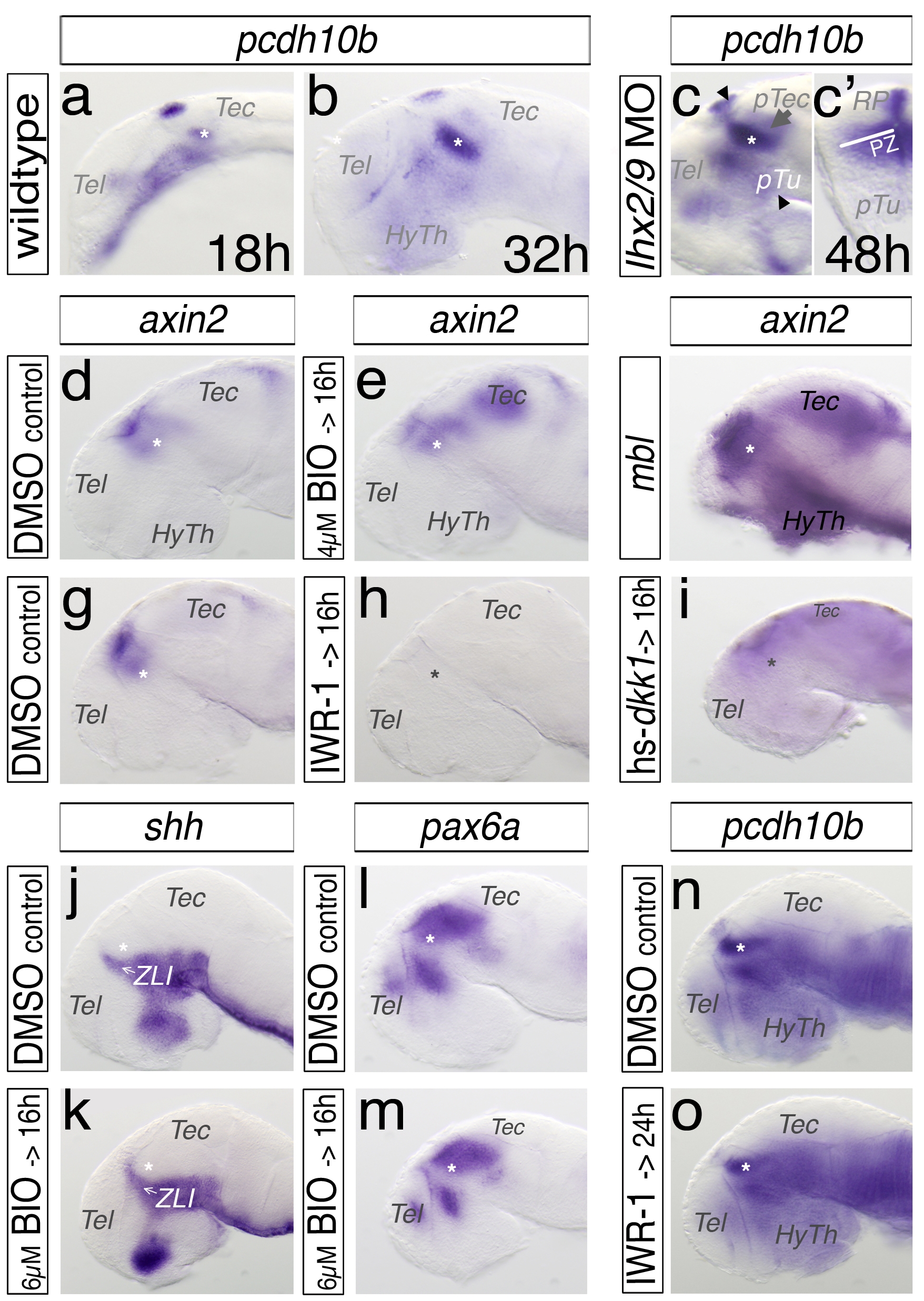
The thalamic expression of protocadherin10b and its regulation. All embryos are analyzed by a single in situ hybridization approach and mounted laterally, with stages indicated, except (c2) shows a cross-section and the left hemisphere is displayed. In the thalamus (asterisk) pcdh10b reveals an onset of expression in segmentation phase (18 hpf), which increases during development (a, b). Knock-down of Lhx2/Lhx9 leads to an expansion of pcdh10b expression into the pretectum (pTec, c), as well as of the ventricular zone (VZ, white bar, c2). Black arrowheads indicate the plane of a cross-section. To validate the efficiency of pharmacological treatment with the Wnt signaling agonist BIO or antagonist IWR-1, we also analyzed under the same conditions the Wnt target gene axin2. Treatment with the Wnt signaling agonist BIO demonstrates an up-regulation of axin2, displayed lateral (d, e). Axin2 expression is upregulated in axin1 mutant embryo masterblind (mbl, f). The treatment of embryos with the Wnt signaling antagonist IWR-1 leads to a loss of axin2 in the diencephalon (g, h). We find a similar reduction of axin2 expression in embryos expressing Dkk1 post-heat-shock at 16 h (i). Treatment of embryos with the Wnt agonist BIO has no effect in the expression of shh or pax6a in the forebrain (j–m). In contrast, embryos treated with the antagonist IWR-1 after endogenous pcdh10b induction between 24 hpf and 48 hpf show no change in pcdh10b expression pattern. HyTh, hypothalamus; pTec, pretectum; pTu, posterior tuberculum; RP, roof plate; Tec, tectum; Tel, telencephalon.