
|
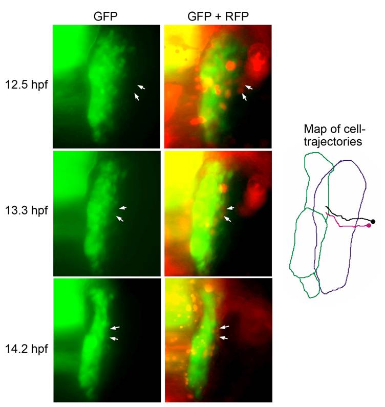
Fig. S2 Recruitment of lateral cells into the pax2a:GFP domain. Representative frames from a movie of a pax2a:GFP transgenic embryo injected with cmv:RFP plasmid DNA. RFP-positive cells originating from a position lateral to the otic/epibranchial domain were tracked as they entered the pax2a:GFP domain and activated expression of GFP. White arrows indicate the positions of two cells with respect to domains of GFP alone or both GFP and RFP. A map of the migration patterns of the two tracked cells is indicated, with the purple and green borders marking the initial and final positions, respectively, of the pax2a:GFP domain.