
|
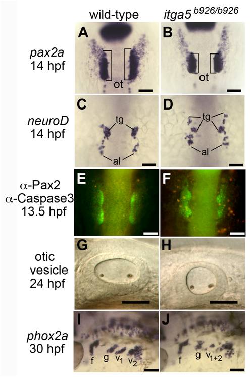
Fig. S1 Abnormal development of posterior placodes in itga5b926/b926 mutants. (A, B) pax2a expression at 14 hpf in the otic/epibranchial domain in a wild-type embryo (A) and itga5 mutant (B). Otic placodes (o, brackets) are indicated. (C, D) neuroD expression at 14 hpf in a control embryo (C) and itga5 morphant (D). Precursors of the trigeminal ganglion (tg) and anterior lateral line (al) are indicated. (E, F) Immunolocalization of Pax2 (green) and Caspase 3 (red) in a wild-type embryo (E) and itga5 mutant (F). (G, H) Otic vesicles at 24hpf in a wild-type embryo (G) and itga5 mutant (H). (I, J) phox2a expression in epibranchial ganglia at 30 hpf in a wild-type embryo (I) and itga5 mutant (J). Facial (f), glossopharyngeal (g), and vagal ganglia (v1+v2) are indicated. A–E are dorsal views with anterior to the top; G–J are lateral views with anterior to the left. Scale bar, 50 μm.