
|
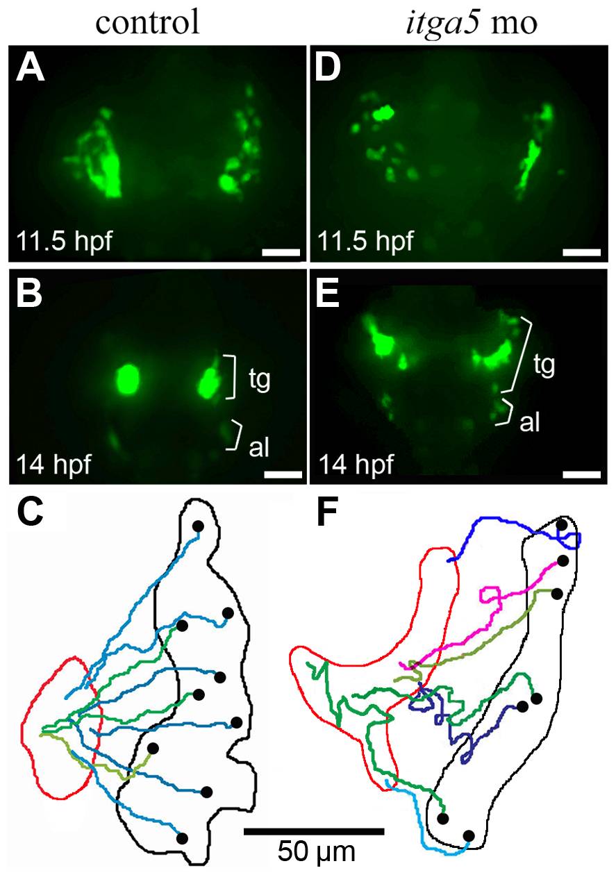
Fig. 5 Convergence of trigeminal precursors is impaired in itga5 morphants.
(A, B, D, E) Images of time-lapse movies showing transgenic neuroD:EGFP expression in the first (11.5 hpf) and final (14 hpf) frames of a control movie (A, B) and itga5 morphant movie (D, E). Positions of precursors of the trigeminal ganglion (tg) and anterior lateral line (al) are indicated. (C, F) Maps showing the trajectories of individual trigeminal precursors in the control embryo (C) and itga5 morphant (F). Black and red boundaries mark the initial and final distribution, respectively, of neuroD:EGFP-positive trigeminal precursors. Black dots represent the initial and final positions, respectively, of individual cells. Images show dorsal views with anterior to the top, and summary figures show the right trigeminal field of each embryo, with lateral to the right. Scale bar, 50 μm.