
|
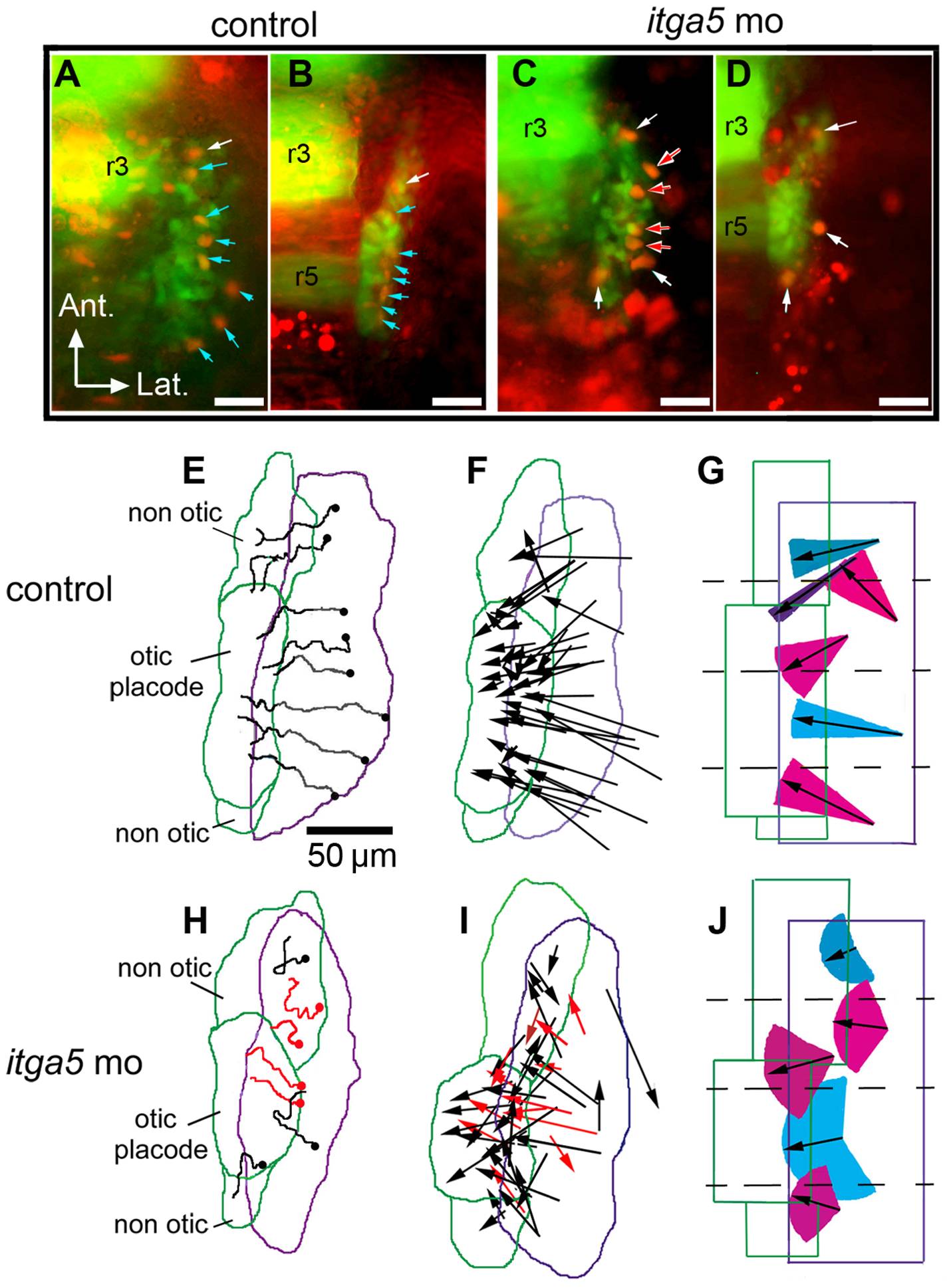
Fig. 2 Otic/epibranchial precursors show aberrant migration in itga5 morphants.
(A–D) Images from time-lapse movies showing transgenic expression of pax2a:GFP (green) and mosaic expression of cmv:RFP (red). The first frame (11.5 hpf) and final frame (14.5 hpf) of a control movie (A, B) and itga5 morphant movie (C, D) are shown. Arrows indicate cells that expressed both GFP and RFP. Blue arrows indicate cells that contributed to the otic domain, and white arrows indicate cells that contributed to non-otic domains. Red arrows indicate cells that lysed during the recording period (C, D). Positions of rhombomeres 3 and 5 (r3, r5) are indicated. (E, H) Maps showing the trajectories of all marked cells in the embryos recorded in A–D. Trajectories in red denote cells that lysed during recording (H). The origins of cell trajectories are marked with dots. The initial and final positions of the pax2a:GFP domain are indicated by purple and green boundaries, respectively. Final positions of the otic placode and non-otic domains are indicated. (F, I) Vector maps showing net displacement of all cells tracked in 5 control movies (F) and 4 itga5 morphant movies (I). Red arrows indicate cells that died during recording (I). (G, J) Summaries of average migration patterns of cells in different quadrants of the pax2a:GFP domain in control embryos (G) and itga5 morphants (J). Arrow length indicates the mean of the net displacement of cells in the indicated region, and colored cones represent the range of angle of net displacement. Quadrants 1 and 2 contained cells contributing to both otic and non-otic domains, which were grouped separately. All images depict the right half of the embryo with lateral to the right and anterior to the top. Scale bar, 50 μm.