
|
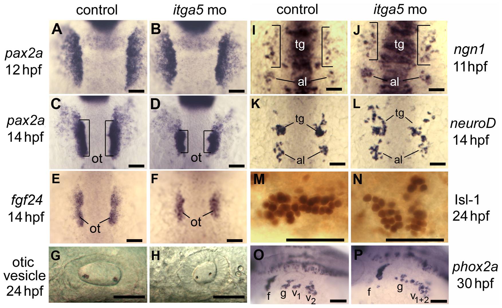
Fig. 1 Knockdown of itga5 impairs morphogenesis of posterior cranial placodes.
(A–D) pax2a expression at 12 and 14 hpf in the otic/epibranchial domain in control embryos (A, C) and itga5 morphants (B, D). Expression is normal at 12 hpf in itga5 morphants but the otic placode (o, brackets) is smaller than normal by 14 hpf. (E, F) fgf24 expression at 14 hpf in a control embryo (E) and itga5 morphant (F). (G, H) Otic vesicles at 24hpf in a control embryo (G) and itga5 morphant (H). (I, J) ngn1 expression at 11 hpf in a control embryo (I) and itga5 morphant (J). (K, L) neuroD expression at 14 hpf in a control embryo (K) and itga5 morphant (L). Precursors of the trigeminal ganglion (tg) and anterior lateral line (al) are indicated. (M, N) Anti-Isl-1 immunostaining at 24 hpf in a control embryo (M) and itga5 morphant (N). (O, P) phox2a expression in epibranchial ganglia at 30 hpf in a control embryo (O) and itga5 morphant (P). Facial (f), glossopharyngeal (g), and vagal ganglia (v1+v2) are indicated. A–E, I–K are dorsal views with anterior to the top; G, H, M–P are lateral views with anterior to the left. Scale bar, 50 μm.