
Image
|
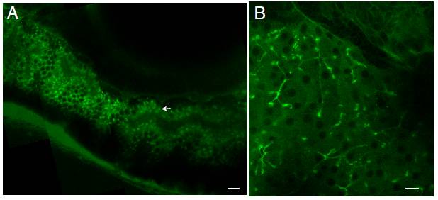
Figure Caption
Fig. S2 The BODIPY fluorophore enters intestinal but not hepatic lipid droplets. A. BODIPY-fed larvae (6 dpf) exhibit fluorescent intestinal lipid droplets (arrow) throughout the enterocytes of their anterior intestine. B. In the liver, BODIPY appears mainly in the hepatic ducts (arrow) and diffusely in the cytoplasm of hepatocytes. Hepatocyte nuclei are indicated (arrowhead). Scale bars = 10μm (n = 3 feeds; 4 larvae imaged per feed).
Acknowledgments
This image is the copyrighted work of the attributed author or publisher, and
ZFIN has permission only to display this image to its users.
Additional permissions should be obtained from the applicable author or publisher of the image.
Reprinted from Developmental Biology, 360(2), Carten, J.D., Bradford, M.K., and Farber, S., Visualizing digestive organ morphology and function using differential fatty acid metabolism in live zebrafish, 276-85, Copyright (2011) with permission from Elsevier. Full text @ Dev. Biol.