
|
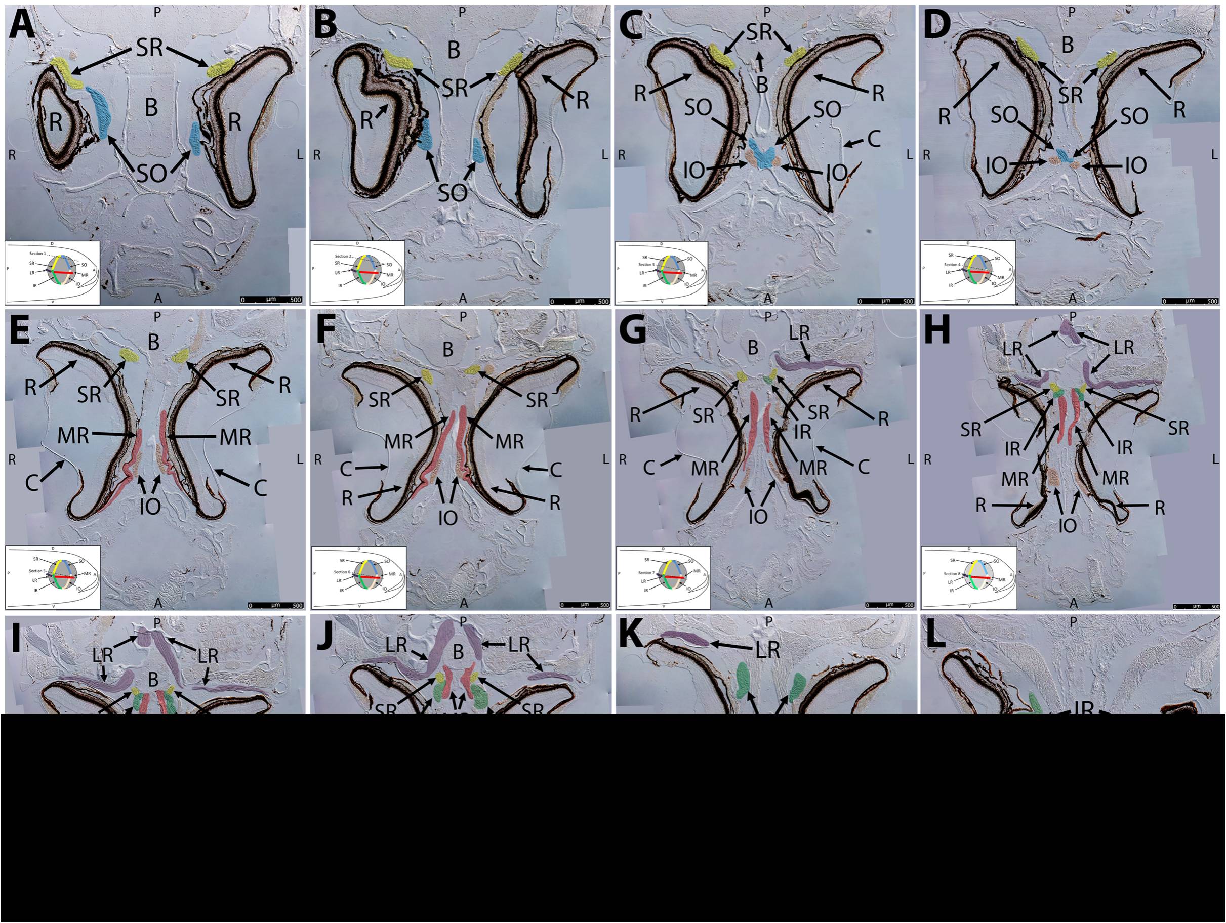
Fig. 4 Key anatomical features within the WT adult zebrafish orbit are highlighted on 12 μm thick coronal sections originally imaged at 200X magnification with DIC prisms to reveal topographical tissue architecture.
Sections proceed in the dorsal (4A) to ventral (4L) direction and show all 6 muscles extending from origin to globe insertion. Please refer to the text for further details. Specific EOMs can be observed on the following figures: Superior oblique (4A–4D). Superior rectus (4A–4J). Inferior Oblique (4C–4L). Inferior rectus (4G–4L). Medial rectus (4E–4J). Lateral rectus (4G–4K). Anterior (A), posterior (P), left (L), and right (R) directions are noted on each frame and a schematic illustrating the specific plane of section is located in the lower left corner. Key features are labeled as follows: B – brain, C – cornea, IO – inferior oblique, IR – inferior rectus, LR – lateral rectus, L – lens, MR – medial rectus, ON – optic nerve, SO – superior oblique, SR – superior rectus, R – retina. For larger versions of the images, please refer to supplemental figures 1A-1L.