
Image
|
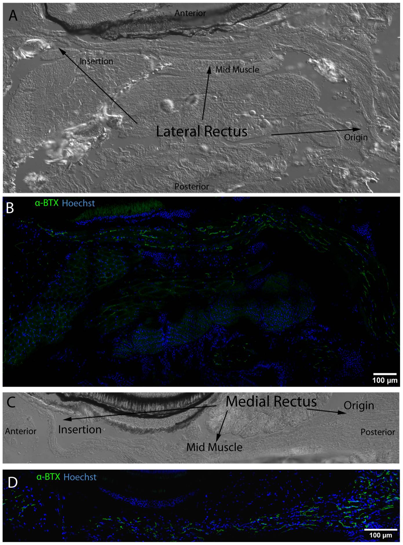
Figure Caption
Fig. 11 NMJ post-synapses labeled with α-BTX (green) reveal even distribution from EOM origin to insertion.
DIC images display full length LR and MR in longitudinal section from origin to insertion (A,C). NMJs (green) labeled with fluorescently conjugated α-BTX are distributed throughout both muscles from origin to insertion and do not appear to be organized into any particular longitudinal pattern across EOM (C,D). Nuclei are stained blue with Hoechst nuclear stain throughout. Mosaics were originally captured using a 63x objective lens.
Acknowledgments
This image is the copyrighted work of the attributed author or publisher, and
ZFIN has permission only to display this image to its users.
Additional permissions should be obtained from the applicable author or publisher of the image.
Full text @ PLoS One