
|
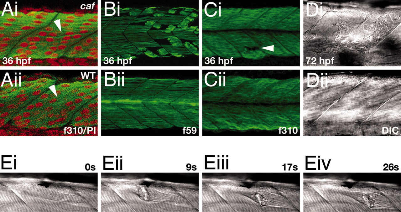
Fig. 1
Initial muscle formation and degeneration in caf embryos. (A) Myoblasts fuse and elongate to form multinucleate fibers (arrowheads) that span the myotome between vertical myosepta. Single confocal scan through epaxial somite at 36 hpf. Green, f310 anti-fast MyHC; red, propidium iodide. (B) Degeneration in the slow layer, which is the first to elongate and fuse. Confocal projection of somite at 36 hpf. f59 anti-slow MyHC. (C) Degeneration in the deeper fast muscle layer. Arrowhead indicates a site of fiber loss. Confocal projection of somite at 36 hpf, f310 anti-fast MyHC. (D) Myotomal lesions at 72 hpf. DIC brightfield image of epaxial somite. (E) Stills from a time-lapse DIC movie showing fiber detachment and retraction in a homozygous caf embryo at 72 hpf, captured by using the anesthetic recovery technique. Time is indicated in seconds. The full movie sequence is available as SI Movie 1. In all images, anterior is to the left.