
Image
|
Figure Caption
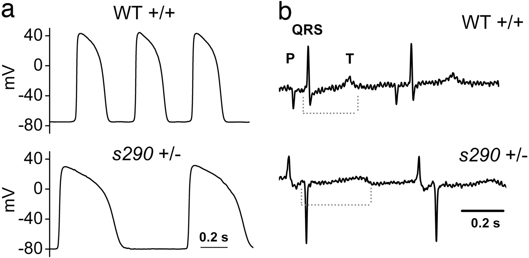
Fig. 5
Heterozygous kcnh2 zebrafish manifest delayed ventricular repolarization. (a) APs recorded from 48 hpf heterozygous ventricle show increased AP duration compared with wild-type (see Results). (b) Representative electrocardiograms recorded from anesthetized, paralyzed wild-type and heterozygous adult zebrafish. Dashed line indicates duration of QT interval. QT corrected for heart rate (see Materials and Methods) was 396 and 467 msec for the exemplar wild-type and heterozygote, respectively.
Figure Data
Acknowledgments
This image is the copyrighted work of the attributed author or publisher, and
ZFIN has permission only to display this image to its users.
Additional permissions should be obtained from the applicable author or publisher of the image.
Full text @ Proc. Natl. Acad. Sci. USA