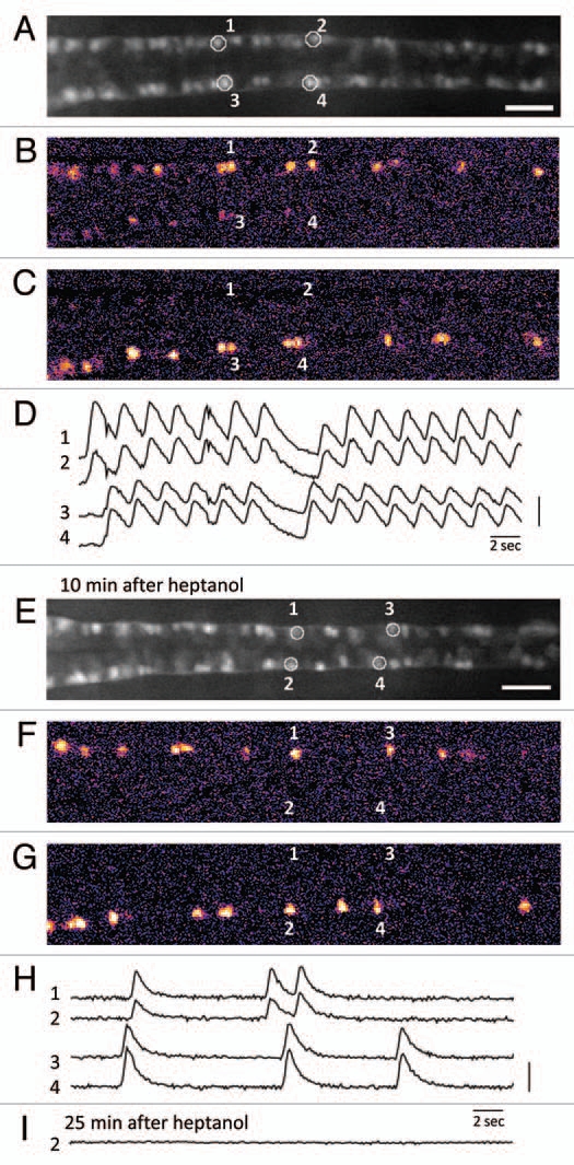
Fig. 1 Calcium imaging of the motor circuit in the zebrafish spinal cord during spontaneous contractions. (A) Expression of GCaMP-HS in the CaP motor neurons in the spinal cord. A dorsal view of the SAIG213A;UA S:GCaMPHS double transgenic embryo embedded in 2% low-melting agarose under a fluorescence microscope and treated with a neuromuscular junction blocker, D-tubocurarine. CaP neurons #1–#4 were used as ROIs (regions of interest). Anterior to the left. Scale bar: 200 µm. (B and C) Calcium signals of the CaP motor neurons with pseudocolors (see also www.youtube.com/watch?v=6y44uxrh7z4). (B) The CaP motor neurons on the right side including ROI-1 and -2 showed increased fluorescence. (C) The CaP motor neurons on the left side including ROI-3 and -4 showed increased fluorescence. (D) The fluorescence changes in the selected CaP motor neurons. 1 and 2 (3 and 4) are activated synchronously, and the right (1 and 2) and left (3 and 4) neurons are activated alternately. A vertical bar indicates (F-resting F)/resting F = 50%. (E) A dorsal view of the SAIG213A;UA S:GCaMPHS double transgenic embryo embedded in 2% low-melting agarose under a fluorescence microscope and treated with a gap junction blocker, heptanol, for 10 min. Anterior to the left. Scale bar: 200 µm. (F and G) Calcium imaging of the CaP motor neurons in the presence of heptanol with pseudocolors (see also www.youtube.com/watch?v=3xhw9D35H5w). (F) The CaP motor neurons on the right side including ROI-1 and -2 showed increased fluorescence. (G) The CaP motor neurons on the left side including ROI-3 and -4 showed increased fluorescence. (H) The fluorescence changes in the selected CaP motor neurons. Synchronized activation is still observed. A vertical bar indicates (F-resting F)/resting F = 100%. (I) The fluorescence changes are not detected 25 min after the heptanol treatment.
Image
Figure Caption
Acknowledgments
This image is the copyrighted work of the attributed author or publisher, and
ZFIN has permission only to display this image to its users.
Additional permissions should be obtained from the applicable author or publisher of the image.
Full text @ Commun. Integr. Biol.

