
Image
|
Figure Caption
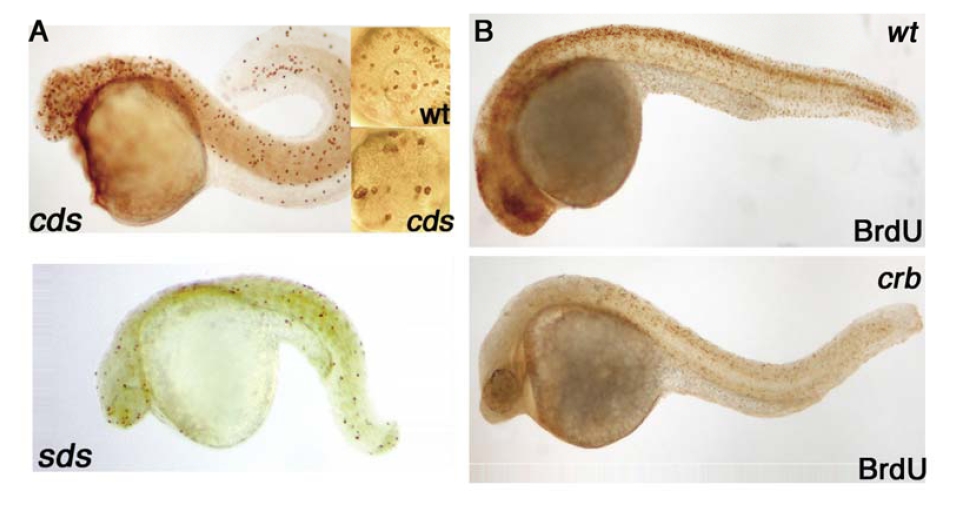
Fig. S5
A cell proliferation screen identified zebrafish cell-cycle mutants. (A) Depiction of the cell proliferation phenotype (by pH3 staining) of two of the eight mutants isolated in the screen: standstill (sds), and cease&desist (cds). For cds, insets represent wild-type (top) and cds mutant (bottom) eyes. All embryos are 36-hpf diploids. (B) Relative levels of cells in S phase as judged by BrdUrd incorporation in crb and wild-type embryos at 28 hpf.
Figure Data
Acknowledgments
This image is the copyrighted work of the attributed author or publisher, and
ZFIN has permission only to display this image to its users.
Additional permissions should be obtained from the applicable author or publisher of the image.
Full text @ Proc. Natl. Acad. Sci. USA