
Image
|
Figure Caption
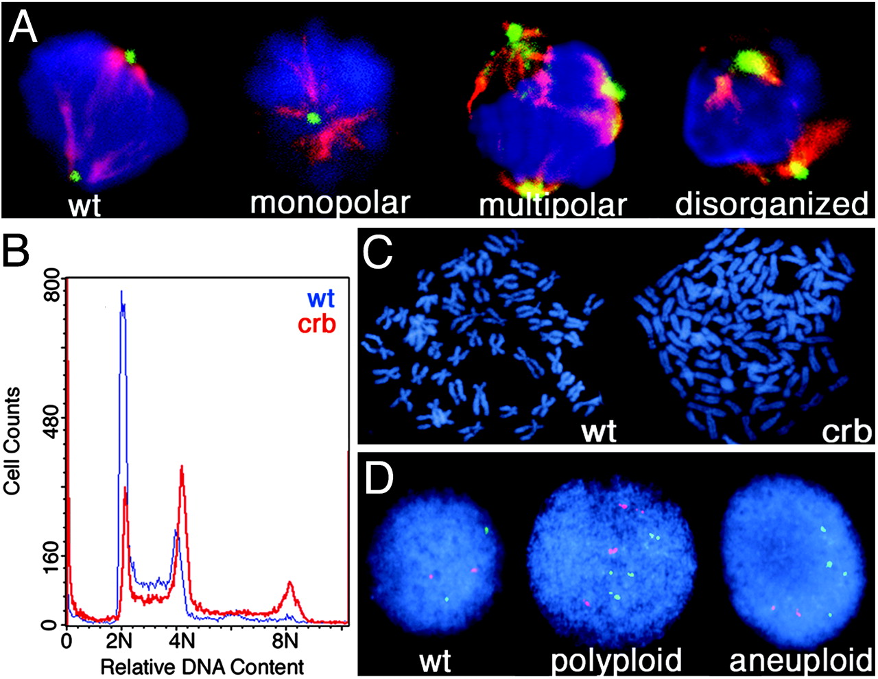
Fig. 3
crb embryos exhibit abnormal mitoses and genomic instability. (A) Mitotic cells shown stained with DAPI (blue), α-tubulin (red), and γ-tubulin (green). (B) DNA content of crb (red) and wild-type (blue) embryos as measured by flow cytometry. (C) Metaphase chromosome spreads from a diploid cell containing 50 chromosomes (Left) and a crb hypertetraploid cell containing 108 chromosomes (Right). (D) FISH analysis on crb mutant embryos with probes for linkage group 2 (red) and linkage group 16 (green) and DAPI counterstaining.
Figure Data
Acknowledgments
This image is the copyrighted work of the attributed author or publisher, and
ZFIN has permission only to display this image to its users.
Additional permissions should be obtained from the applicable author or publisher of the image.
Full text @ Proc. Natl. Acad. Sci. USA