
|
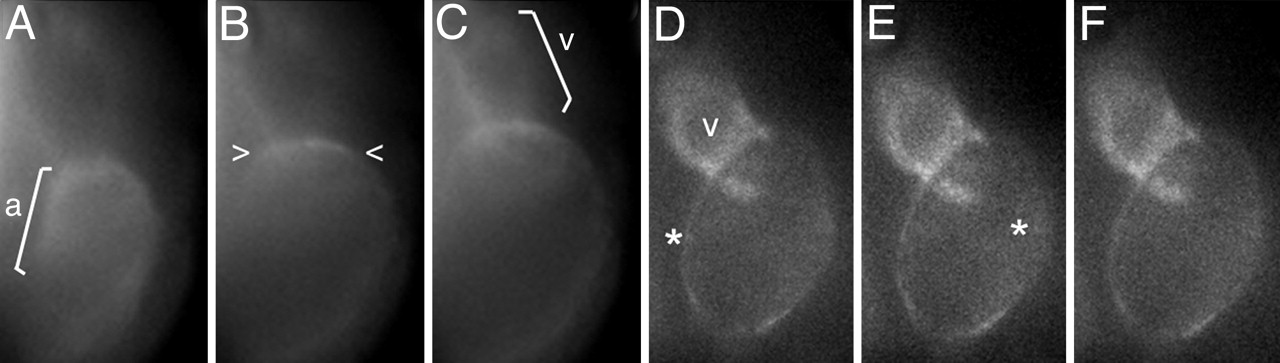
Fig. 5
NCX1h mutation leads to calcium overload in the tre ventricle. Analysis of intracellular calcium levels in 48 hpf embryos by calcium orange fluorescent dye. Selected sequential frames at H70-msec intervals are shown. (A–D) In wild-type embryos, Ca2+ entry into cardiomyocytes, denoted by dye, sweeps across the heart in a wave (brackets) correlated with the sequential contraction of the chambers. (E–H) In tre mutants, calcium orange levels in the ventricle remain static and appear elevated relative to the atrium, suggesting calcium overload in the ventricle. Calcium signals corresponding to one or a few adjoining cells briefly fluoresce in the fibrillating atrium (see faint signals to right of *). Image is anterior to the top. a, atrium; v, ventricle.