
Fig. 1
- ID
- ZDB-IMAGE-111207-11
- Genes
- Publication
- Baker et al., 2008 - Direct and indirect roles for Nodal signaling in two axis conversions during asymmetric morphogenesis of the zebrafish heart
- All Figures
- Figures for Baker et al., 2008
|
Fig. 1
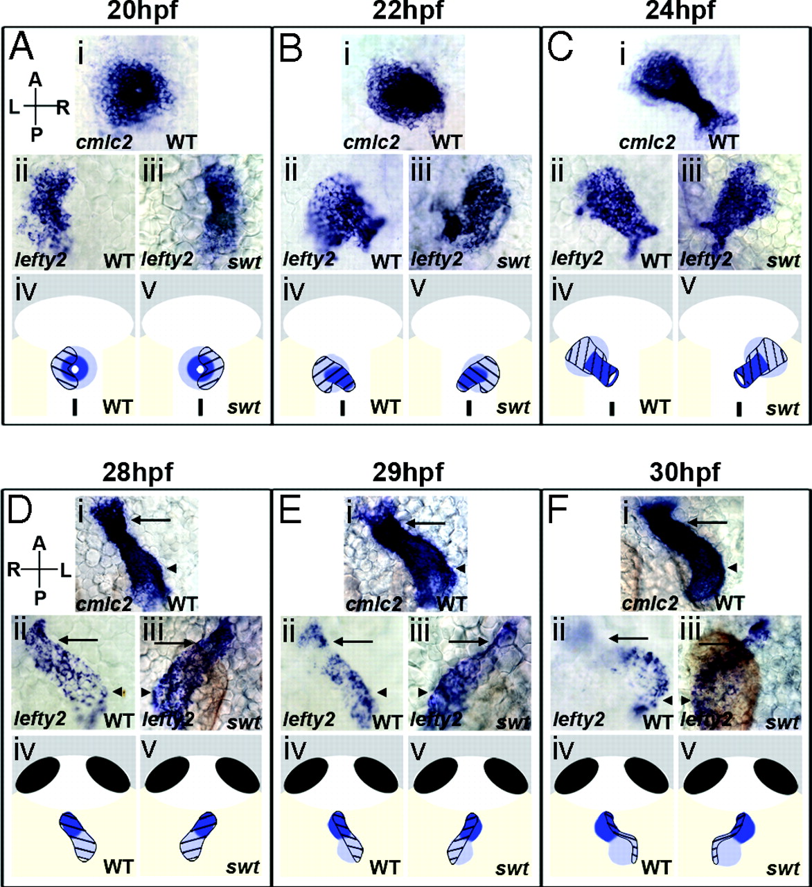
lefty2 expression temporally correlates with establishment of morphological asymmetries in the heart. Dorsal (A–C) and ventral (D–F) views of the heart from 20–30 hpf. Arrows indicate the ventricle and arrowheads the atrium (Di–Fiii). Cardiac cells were visualized by RNA in situ hybridization for cmlc2, expressed in all myocardial cells, and lefty2. In WT, lefty2 expression is restricted to the left myocardium at 20 hpf (Aii), but extends symmetrically along the length of the left-jogged heart by 22–28 hpf (Bii, Cii, and Dii). A swt morphant exhibits reversed lefty2 expression in the right myocardium (Aiii) at 20 hpf. At 22–28 hpf, lefty2 is expressed along the length of the right-jogged heart in swt morphants (Biii, Ciii, and Diii). At 28–30 hpf, lefty2 expression becomes restricted to the left side of the heart in WT (Eii) and is expressed exclusively along the left margin of the looping heart at 30 hpf (Fii). This process is reversed in swt morphants at 29 hpf. In this embryo, lefty2 expression is still visible across the atrium (Eiii, arrowhead) but it restricted to the right side of the ventricle (Eiii, arrow). Right-sided localization is more pronounced by 30 hpf, with lefty2 expression restricted to the right in both chambers (Fiii). The diagrams show atrial cells (light blue), ventricular cells (dark blue), localization of lefty2 (black hatchmarks). L, left; R, right; A, anterior; P, posterior.