
|
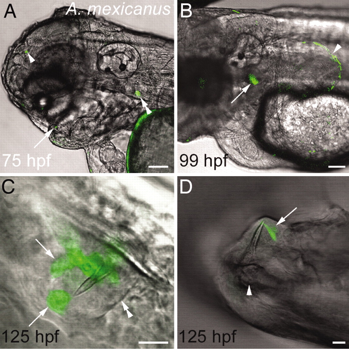
Fig. 4
The zebrafish dlx2b:GFP reporter drives expression in developing A. mexicanus oral teeth, pharyngeal teeth, and pectoral fins. (A) Lateral view of the head at 75 hpf with expression in an oral tooth germ (arrow), upper pharyngeal tooth germ (double arrowhead), and a small ectopic location (arrowhead). (B) Lateral view of pharyngeal tooth (arrow) and pectoral fin (arrowhead) expression at 99 hpf. (C) Oral tooth forming in the upper jaw adjacent to the developing premaxillary bone at 125 hpf. The boundary of the dental epithelium can be seen on one side (double arrowhead) and two groups of dlx2b:GFP reporter expressing cells on the other (arrows). (D) GFP expression in dental epithelial cells (arrow) surrounding a mineralized tooth attached to the dentary bone of the lower jaw (arrowhead) at 125 hpf. (Scale bars: A and B, 100 μm; C and D, 10 μm.)