
|
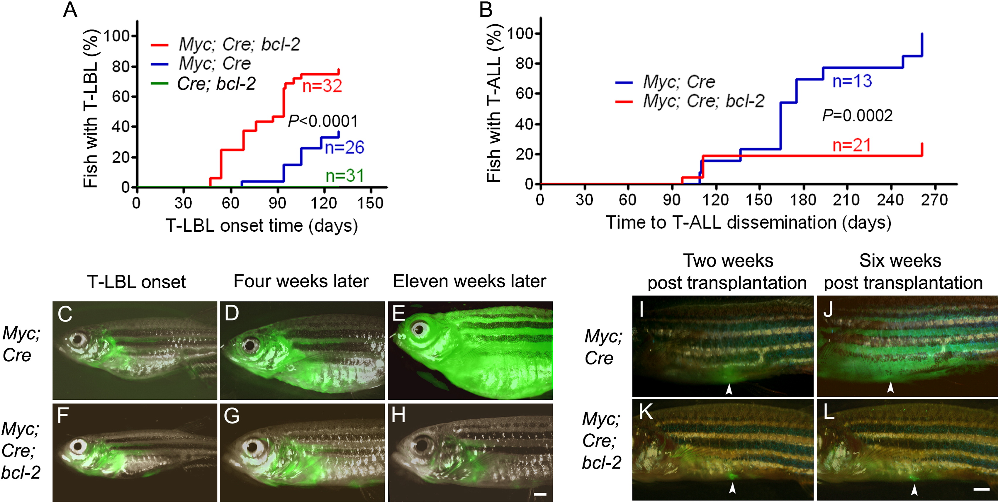
Fig. 1 Bcl-2 Promotes Onset but Inhibits the Progression of Myc-Induced T-LBL in Zebrafish
(A) Rate of tumor onset in three transgenic zebrafish lines: hsp70-Cre;rag2-EGFP-bcl-2 (Cre;bcl-2) double-transgenic fish (n = 31; green line), rag2-LDL-EGFP-Myc;hsp70-Cre (Myc;Cre) double-transgenic fish (n = 26; blue line), and rag2-LDL-EGFP-Myc;hsp70-Cre;rag2-EGFP-bcl-2 (Myc;Cre;bcl-2) triple-transgenic fish (n = 32; red line).
(B) Rate of T-LBL progression to T-ALL in Myc;Cre (n = 13; blue line) versus Myc;Cre;bcl-2 (n = 21; red line) transgenic fish.
(C–H) Localized GFP-labeled tumors first arose as T-LBL in Myc;Cre (C; 112-day) and Myc;Cre;bcl-2 (F; 119-day) transgenic fish; widespread dissemination leading to leukemia was seen within 11 weeks after T-LBL onset in Myc;Cre fish (D and ,E), but not in Myc;Cre;bcl-2 triple transgenics (G and H).
(I–L) GFP-positive T-LBL tumor cells (n = 5 per group) transplanted into the peritoneum of irradiated wild-type hosts. Tumor cells from the Myc;Cre double-transgenic fish disseminated rapidly (I–J), while those from the Myc;Cre;bcl-2 triple-transgenics remained localized (K and L). Scale bar for (C)–(H) and (I)–(L), 1 mm.
See also Figure S1.
Reprinted from Cancer Cell, 18(4), Feng, H., Stachura, D.L., White, R.M., Gutierrez, A., Zhang, L., Sanda, T., Jette, C.A., Testa, J.R., Neuberg, D.S., Langenau, D.M., Kutok, J.L., Zon, L.I., Traver, D., Fleming, M.D., Kanki, J.P., and Look, A.T., T-lymphoblastic lymphoma cells express high levels of BCL2, S1P1, and ICAM1, leading to a blockade of tumor cell intravasation, 353-366, Copyright (2010) with permission from Elsevier. Full text @ Cancer Cell