
|
Fig. 3 Zebrafish Lymphoblasts Overexpressing Myc and Bcl-2 Undergo Autophagy
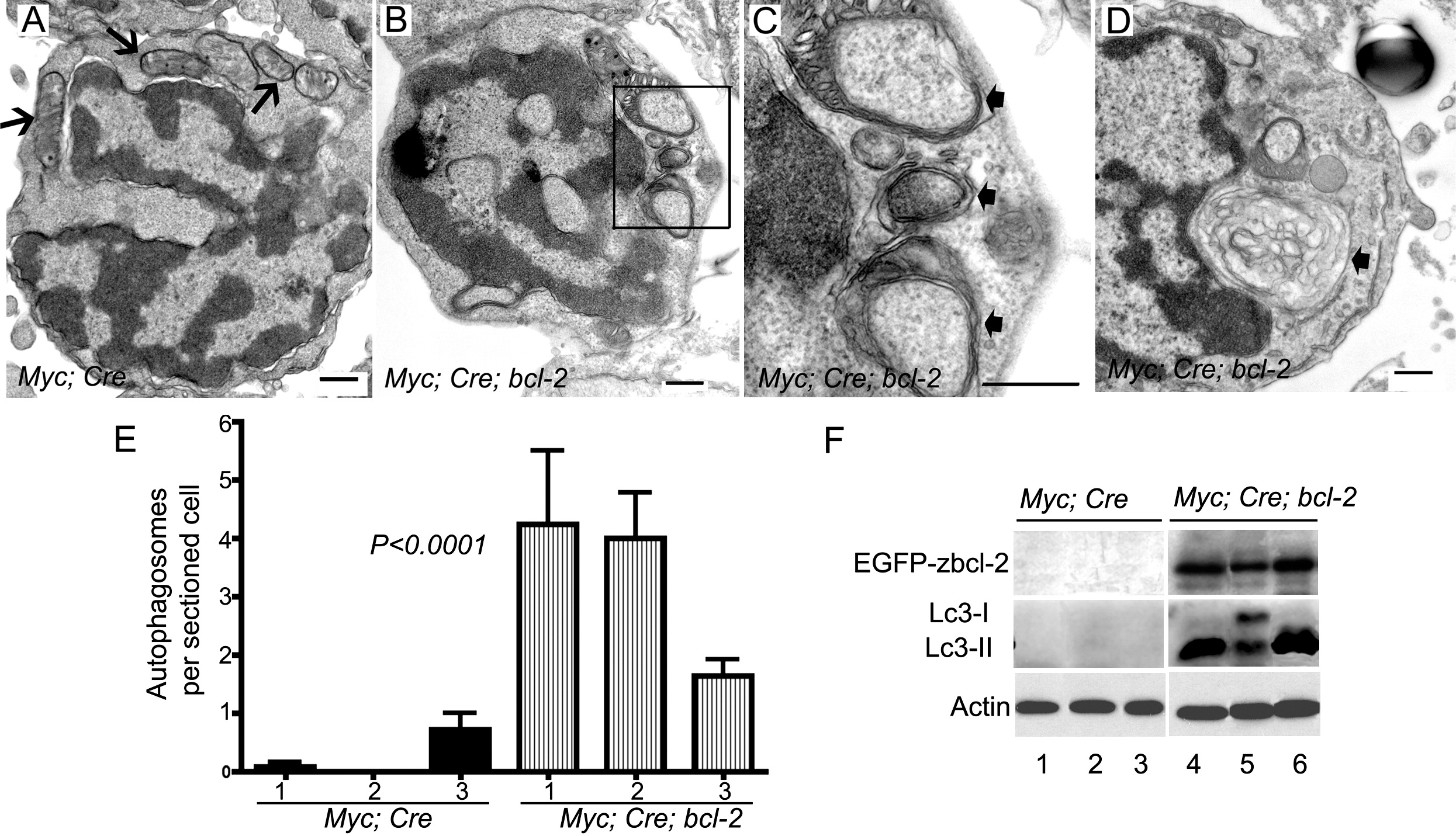
(A) Electron microscopic analysis rarely identified autophagosomes in tumor cells from Myc;Cre transgenic fish. Mitochondria are indicated by arrows.
(B–D) Thymic lymphoblasts from Myc;Cre;bcl-2 triple-transgenics show prominent autophagosomes/autolysophagosomes. (C) is a magnified view of (B) (box). Arrows indicate double-membrane autophagosomes containing cytoplasm and cytoplasmic organelles. An autolysophagosome is shown in (D) (arrow).
(E) Quantification of autophagosomes and autolysophagosomes in Myc;Cre (solid bars) and Myc;Cre;bcl-2 (hatched bars) tumor cells were harvested from three individual fish. From 9 to 15 different cells from each fish were sectioned and analyzed. Mean ± SD results from three individual fish are shown.
(F) Western blot analyses of the protein levels of EGFP-zbcl-2, Lc3-I, and Lc3-II in three individual Myc;Cre and Myc;Cre;bcl-2 transgenic fish. Actin was used as a loading control in each lane.
Scale bars for (A)–(D), 500 nm.
See also Figure S3.
Reprinted from Cancer Cell, 18(4), Feng, H., Stachura, D.L., White, R.M., Gutierrez, A., Zhang, L., Sanda, T., Jette, C.A., Testa, J.R., Neuberg, D.S., Langenau, D.M., Kutok, J.L., Zon, L.I., Traver, D., Fleming, M.D., Kanki, J.P., and Look, A.T., T-lymphoblastic lymphoma cells express high levels of BCL2, S1P1, and ICAM1, leading to a blockade of tumor cell intravasation, 353-366, Copyright (2010) with permission from Elsevier. Full text @ Cancer Cell