
|
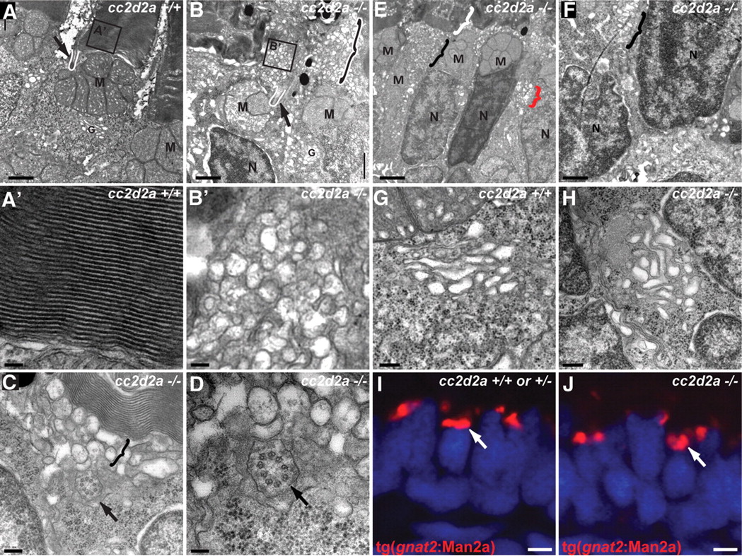
Fig. 6 Loss of Cc2d2a leads to vesicle accumulation in photoreceptors. (A–H) Transmission electron microscopy images of 5 d.p.f. cc2d2a+/+ (A–A′ and G) and cc2d2a-/- (B–F and H) photoreceptors. (A) Low-power image of the inner segment and the base of the outer segment in cc2d2a+/+ photoreceptors showing the mitochondrial cluster M, the connecting cilium (arrow and white line) and the base of the outer segment (A′), with neatly stacked membranes [high power view (A′)]. (B) Low-power image of cc2d2a-/- photoreceptors with normal mitochondrial clusters M and nuclei N, but massive accumulation of vesicles around a normal-appearing connecting cilium [arrow and white line in (B)]. The black and white bracket highlights an outer segment replaced by vesicles. (B′) High-power image of the vesicles boxed in (B). (C and D) Higher power images of the accumulating vesicles (bracket) just below partially stacked membranes. (D) Cross-section through a connecting cilium (arrow) with nine normal appearing microtubule doublets. (E) Lower power image of a different region showing moderate vesicle accumulation (black bracket) below a recognizable outer segment (white bracket). Note the presence of vesicles lateral to the nucleus (red bracket). (F) Higher power image of the basal portion of a photoreceptor showing vesicle accumulations (bracket) between nuclei. (G and H) High-power images of the Golgi apparatus in wild-type (G) and cc2d2a-/- (H) photoreceptors. (I and J) Cryosections of transient transgenic cc2d2a+/+ (I) and cc2d2a-/- (J) embryos expressing the Golgi specific transgene tg(gnat2:Man2a)-RFP (red) under the transducin promoter control. Scale bars are 1 µm in (A–B), 100 nm in (A′ and B′), 200 nm in (C), 100 nm in (D), 2 µm in (E), 1 µm in (F), 500 nm in (G and H) and 2 µm in (I and J). M, mitochondria; N, nuclei; G, Golgi.