
Fig. 5
- ID
- ZDB-IMAGE-111201-54
- Genes
- Antibodies
- Source
- Figures for Bachmann-Gagescu et al., 2011
|
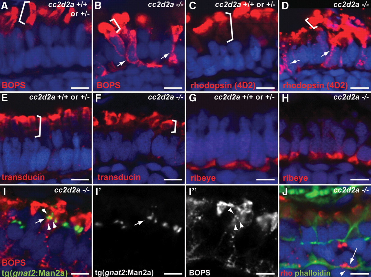
Fig. 5 Selective trafficking defects in cc2d2a-/- photoreceptors. (A and B) BOPS localization (BOPS antibody, red) in wild-type (A) and cc2d2a-/- (B) photoreceptors. (C and D) Rhodopsin localization (4D2 antibody, red) in wild-type (C) and cc2d2a-/- (D) photoreceptors. Outer segments are indicated by brackets. Mislocalized photopigment (opsin) is indicated by white arrows. (E and F) Transducin (red) localization by immunofluorescence is restricted to the outer segments (indicated by brackets) in both cc2d2a+/+ and cc2d2a-/- photoreceptors. (G and H) Ribeye (red) localization at the synapse is indistinguishable between cc2d2a+/+ and cc2d2a-/- photoreceptors. (I-I′′) BOPS staining [red in (I)] and Golgi expression of tg(gnat2:Man2a-RFP, green) do not significantly overlap [see arrowheads in (I) and (I′′) for BOPS and arrow in (I) and (I′) for Golgi]. (J) Rhodopsin (4D2 antibody, red, arrow) is mislocalized between the nucleus (DAPI, blue) and the subcortical actin network stained with phalloidin (green, arrowhead). All images are single-confocal sections of 5 d.p.f. cryosections. Scale bars are 4 µm in all panels.