
Fig. S5
- ID
- ZDB-IMAGE-111201-52
- Publication
- Mo et al., 2011 - Both pre- and postsynaptic activity of nsf prevents degeneration of hair-cell synapses
- All Figures
- Figures for Mo et al., 2011
|
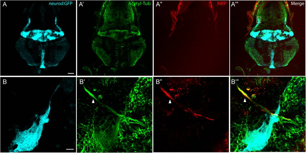
Fig. S5 Myelin Basic Protein expresses in peripheral nerves, but not in the CNS. A-A′′′, A representative top-down projection of a zebrafish brain at 5 dpf labeled by GFP in the TgBAC(neurod:EGFP)nl1 background (A, light blue), antibodies against acetylated Tublin (Acetyl Tub, A′, green) and Myelin Basic Protein (MBP, A′′, red). MBP protein was not detected in the CNS. Scale bar: 100 μm. B-B′′′, Close up of the boxed region in panel A′′′. Although MBP fluorescence was associated with nerve fibers from the anterior lateral line ganglion (arrow head), there was no labeling of MBP in nerve fibers in the cerebellar region (highlighted by GFP expression). Scale bar: 10 μm.