
Fig. S4
- ID
- ZDB-IMAGE-111201-51
- Publication
- Mo et al., 2011 - Both pre- and postsynaptic activity of nsf prevents degeneration of hair-cell synapses
- All Figures
- Figures for Mo et al., 2011
|
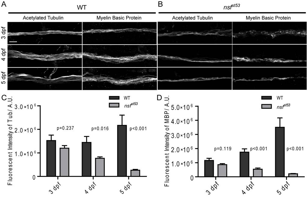
Fig. S4 Decreased labeling of acetylated Tubulin and Myelin Basic Protein in nsfst53 mutants. A, Antibodies against acetylated Tubulin were used to label lateral line nerves in wild-type and nsfst53 mutant larvae at 3, 4 and 5 dpf. B, Anti-Myelin Basic Protein antibody labeled the myelin sheath of the lateral line nerve in both wild-type and nsfst53 mutants (3 to 5 dpf). C, The fluorescent intensity of acetylated Tubulin labeling increased over time in wild-type larvae (3 dpf, 1523307±233786; 4dpf, 1445077±252144; 5 dpf, 2170483±434340), but decreased in nsfst53 mutants (3 dpf, 1201135±110419; 4dpf, 770785±58768; 5 dpf, 264018±35511). D, The fluorescent labeling of Myelin Basic Protein displayed a dramatic increase in wild-type (3 dpf, 1155025±158382; 4dpf, 1746149±232916; 5 dpf, 3501120±670992), but significantly decreased in nsfst53 mutants (3 dpf, 860546±74798; 4dpf, 538577±85907; 5 dpf, 212506±24043). Scale bar: 10 μm; z-projection of 5 confocal planes (1 μm each).