
Fig. S3
- ID
- ZDB-IMAGE-111201-50
- Publication
- Mo et al., 2011 - Both pre- and postsynaptic activity of nsf prevents degeneration of hair-cell synapses
- All Figures
- Figures for Mo et al., 2011
|
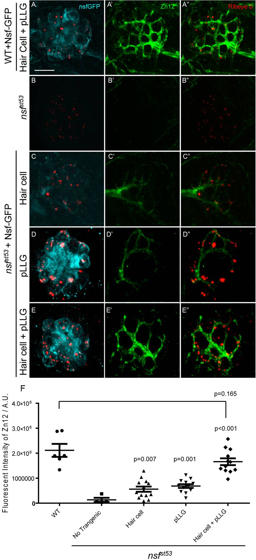
Fig. S3 Rescue of afferent innervation by double transgenic expression of Nsf-GFP in nsfst53 mutants. A-E′′, Top-down views of the first lateral line neuromast (5 dpf) from wild-type (A), nsfst53 mutants (B), and nsfst53 mutants with Nsf-GFP expressed in hair cells (C), pLLG (D), or in both hair cells and pLLG (E). Shown is immunolabeling with antibodies against GFP (light blue), Zn12 (green), and Ribeye b (red). Scale bar: 10 μm. F, The total intensity of Zn12 antibody labeling per neuromast was quantified in wild-type (2.113e6±259343, n = 6),nsfst53 mutants (133379±80303, n = 4), and nsfst53 mutants with Nsf-GFP rescued in hair cells (559662±104829, n = 13), pLLG (684537±75475, n = 12), or both hair cells and pLLG (1.659e6±136058, n = 12).The p-values were generated comparing the data from the nsfst53 mutant to each transgenic mutant line.