
IMAGE
Fig. S1
- ID
- ZDB-IMAGE-111201-49
- Publication
- Mo et al., 2011 - Both pre- and postsynaptic activity of nsf prevents degeneration of hair-cell synapses
- All Figures
- Figures for Mo et al., 2011
Image
|
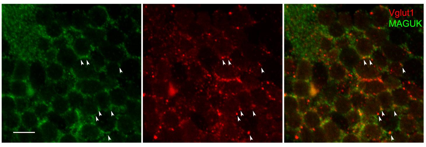
Figure Caption
Fig. S1 Glutamatergic synapses in the cerebellum. Magnified images of the boxed region in Figure 4A′ showed fluorescent signals from Vglut1 (red) and MAGUK (green) antibodies in Purkinje cells of the cerebellar region. Although MAGUK antibody labels both cell body and postsynaptic density in these neurons, it is possible to observe juxtaposition of MAGUK densities next to Vglut1 labeled presynaptic terminals (arrow heads). Scale bar is 10 μm.
Acknowledgments
This image is the copyrighted work of the attributed author or publisher, and
ZFIN has permission only to display this image to its users.
Additional permissions should be obtained from the applicable author or publisher of the image.
Full text @ PLoS One