
Fig. 1
- ID
- ZDB-IMAGE-111201-41
- Genes
- Antibodies
- Publication
- Mo et al., 2011 - Both pre- and postsynaptic activity of nsf prevents degeneration of hair-cell synapses
- All Figures
- Figures for Mo et al., 2011
|
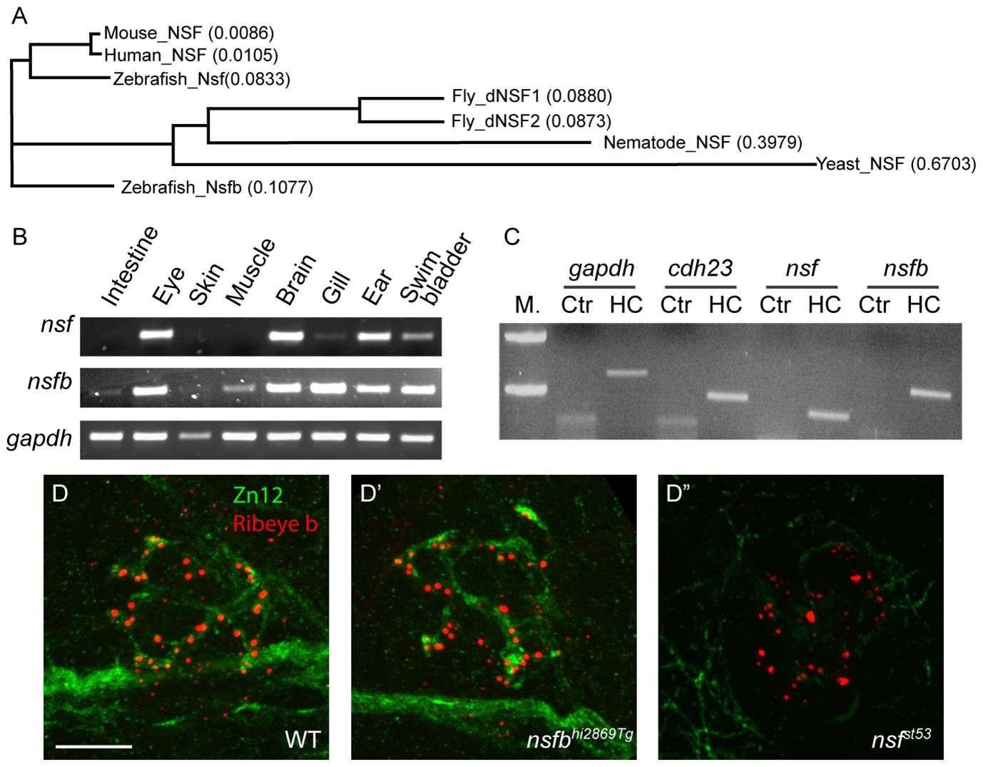
Fig. 1 Expression of nsf and nsfb in zebrafish, and defective innervation of hair cells in nsf mutants.
A, Protein sequences of NSF from yeast, nematode, fly, zebrafish, mouse and human were aligned using ClustalW to generate a phylogenic tree. The total amino acid substitutions of a specific protein are proportional to the length of each branch. The substitute rates of single amino acid were numbered in brackets after each protein. B, Detection of nsf and nsfb transcripts in adult zebrafish tissues by RT-PCR. C, RT-PCR of neuromasts isolated from 5 dpf larvae. cdh23 was a positive control as a neuromast-specific gene. M. = Marker/DNA ladder, Ctr = Control with no reverse transcriptase, HC = Hair cell. D, Larvae (4 dpf) labeled with nerve fiber-specific Zn12 (green), and anti-Ribeye b antibodies (red). The merged images showed the innervation of the first lateral line neuromast (L1) by posterior lateral line neurons. Scale bar, 10 μm. The position of the specimen is dorsal up and anterior to the left, and each image is a projection of 10 optical sections (1 μm each). Scale bar is 10 μm.