
Fig. 3
- ID
- ZDB-IMAGE-111129-23
- Publication
- Goudevenou et al., 2011 - Def6 Is Required for Convergent Extension Movements during Zebrafish Gastrulation Downstream of Wnt5b Signaling
- All Figures
- Figures for Goudevenou et al., 2011
|
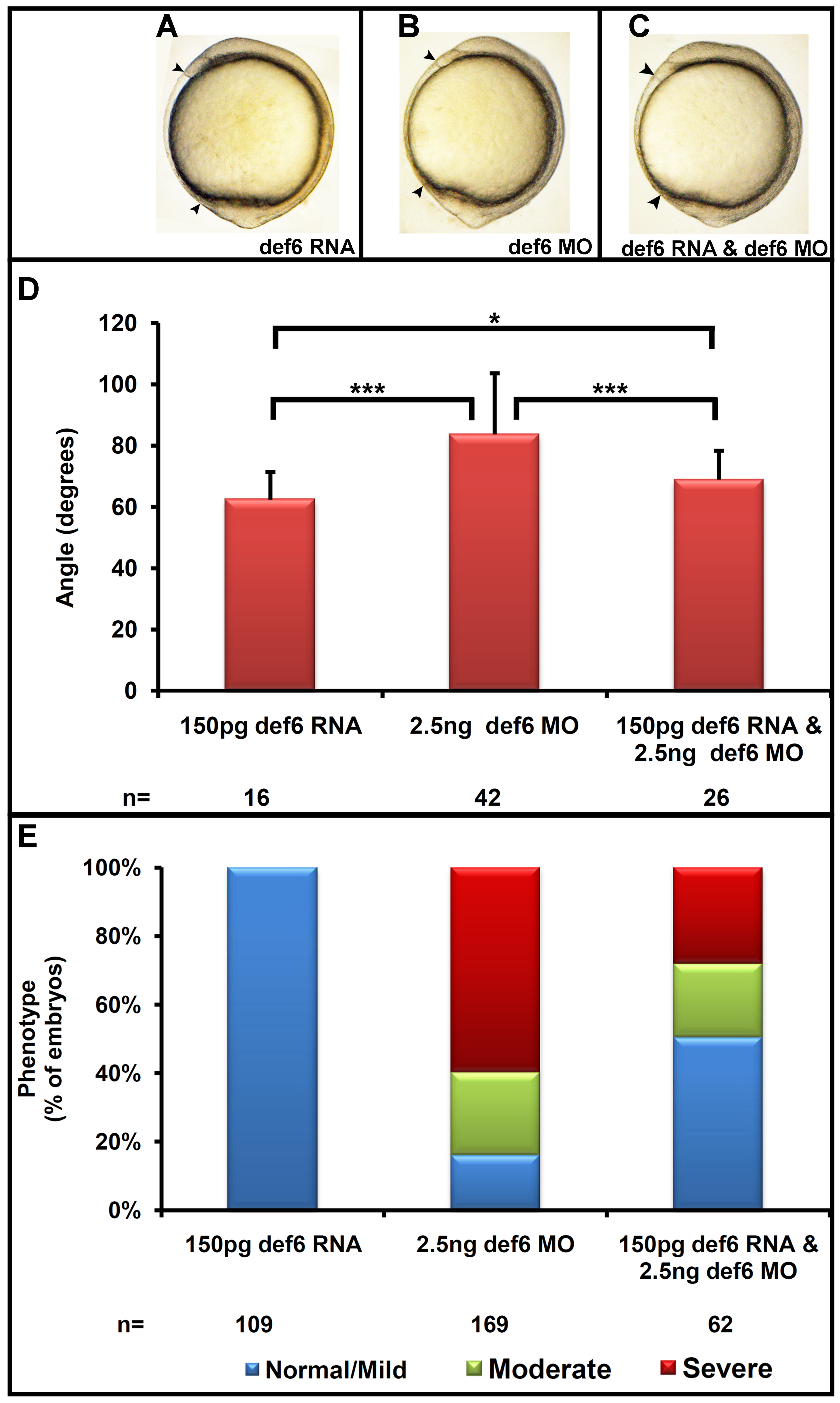
Fig. 3
Def6 RNA rescues the def6 MO-induced phenotype.
(A–C) Def6 splice MO was injected alone (2.5 ng) or together with def6 RNA (150 pg). As a control, def6 RNA was also injected alone (150 pg). Embryos are shown at the 1-somite stage. (D) The angle between the anterior- and posterior-most embryonic structures was measured in at least 20 embryos and the average angle is shown on the graph in degrees. ANOVA single factor and two-tailed Student′s t-tests showed a significant (p<0.001; three asterisks) increase in the angle after injection of 2.5 ng def6 MO and a significant (p<0.001; three asterisks) decrease in the angle after addition of def6 RNA. The angle measured between injected controls and rescued embryos was also statistically significant (p<0.05; one asterisk), suggestive of a partial rescue. (E) The phenotypes of the embryos from three independent experiments were scored at 3 dpf and the percentages of normal/mild (blue bar), moderate (green bar) and severe (red bar) morphology are shown. Representative images of embryos are shown in Figure 2 panel E.