
|
Fig. S2
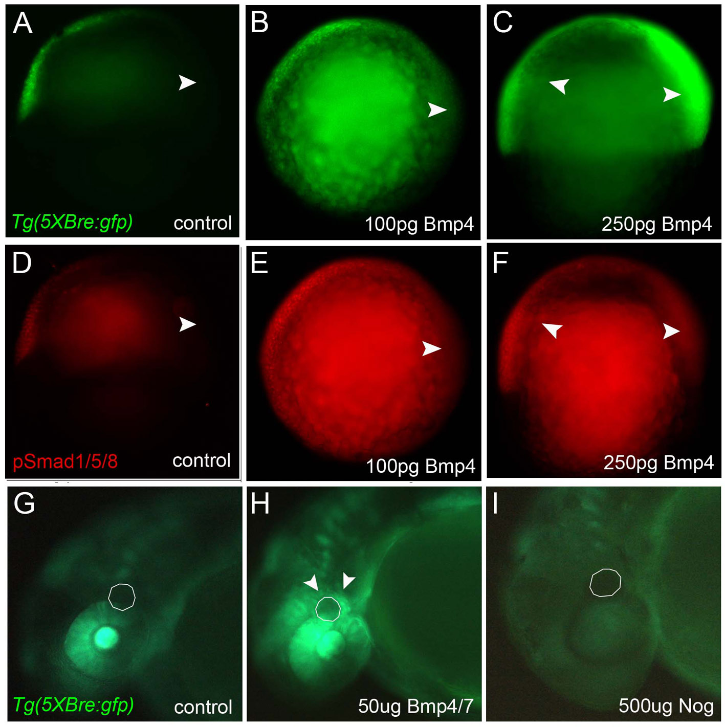
BMP signaling is sufficient and necessary for Tg(Bre:GFP) expression. (A-C) Whole-mount immunochemical staining for GFP protein at early gastrula stages, lateral views, ventral towards the left. (D-F) Whole-mount immunochemical staining for pSmad1/5/8 protein at early gastrula stages, lateral views, ventral towards the left. (A,D) Uninjected controls. (B,E) Embryos microinjected at the one-cell stage with 100 pg xBmp4 mRNA. (C,F) Embryos injected with 250 pg xBmp4. (G-I) Whole-mount immunochemical staining for GFP protein at 30 hpf, lateral views of the head, anterior towards the left. (G) Control embryo in which a DMSO-soaked bead (dashed line) was implanted behind the eye at 24 hpf. (H) Increased GFP surrounding a similarly implanted bead soaked in 10 µg/µl human recombinant BMP4/7. (I) Loss of GFP in an embryo implanted with a bead soaked in 500 μg/μl NOG. Abbreviations: cn, commissural neurons; D, dorsal; de, dorsal eye; m, margin; pa, pharyngeal arches; V, ventral.