
|
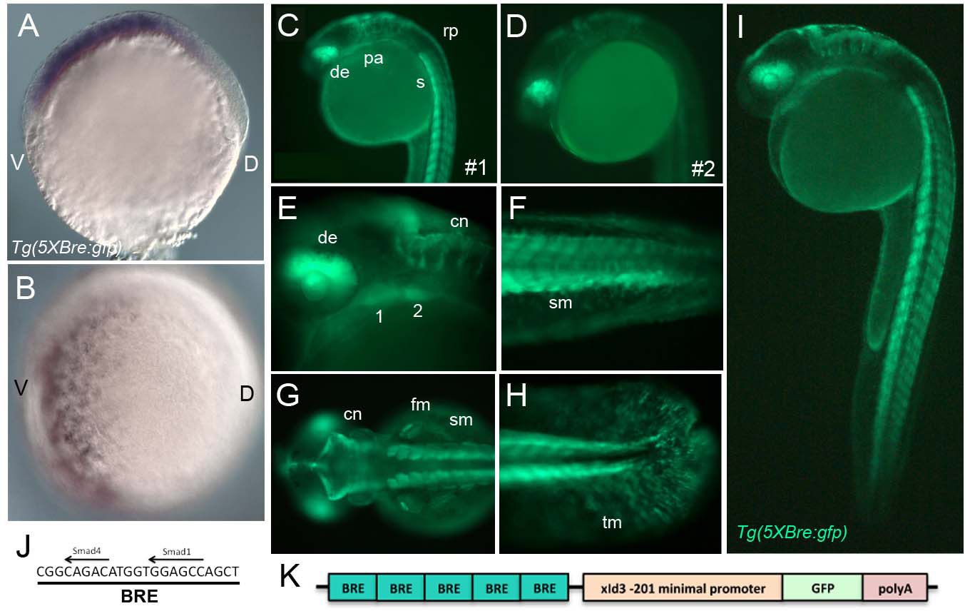
Fig. S1
Tg(Bre:GFP) expression. (A,B) Whole-mount in situ hybridization for GFP mRNA at early gastrula stage (6 hpf), lateral (A) and ventral (B) views, dorsal towards the right. (C-I) Live images of Tg(Bre:GFP) expression, lateral views (except G, which is a dorsal view), anterior to the left. (C,D) Two stable Tg(Bre:GFP) transgenic lines (1 and 2), derived from independent insertion events, show similar expression patterns. (J) The BRE sequence includes putative Smad1- and Smad4-binding sites separated by five bases. (K) The Tg(Bre:GFP) transgenic construct includes five tandem Bres (blue-green), a minimal XId3 promoter (orange), GFP (green) and an SV40 polyA tail (red). 1, mandibular arch; 2, hyoid arch; cn, commissural neurons; de, dorsal eye; fm, fin muscle; pa, pharyngeal arches; sm, somite muscle; tm, tail mesenchyme.