
|
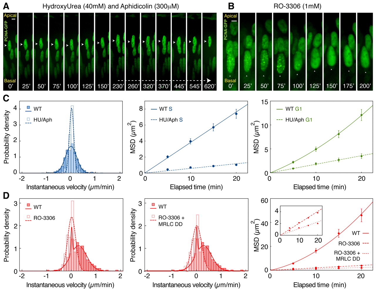
Fig. 5 Cell cycle continuation is required for directed nuclear motion. Time-lapse imaging of nuclei blocked with pharmacological inhibitors of specific cell cycle phases. (A) Kymograph of retinal nucleus (arrowhead) in the presence of the S/G2 inhibitors HU and Aphidicolin, entering but remaining in S phase. (B) Kymograph of a retinal nucleus (arrowhead) in the presence of the G2/M inhibitor RO-3306, entering G2 but not migrating towards the apical surface. (C) Velocity distribution and MSD analysis confirm the absence of directed motion during S phase and a marked reduction in the effective displacement of treated nuclei. The MSD during G1 in treated zebrafish embryos increases linearly with elapsed time, in contrast to wild-type nuclei. (D) Velocity distribution and MSD analysis show the absence of directed motion in RO-3306-treated embryos. This effect cannot be rescued by co-expression of constitutively active Myosin regulatory light chain (MRLC-DD). Error bars indicate s.e.m. Scale bars: 5 μm.