
Image
|
Figure Caption
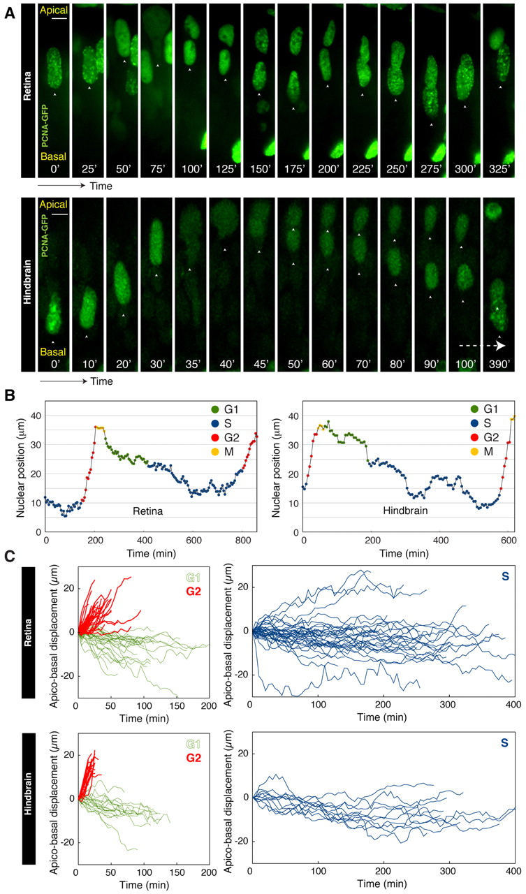
Fig. 2 Modes of nuclear migration tied to specific cell cycle phases. (A) Time-lapse imaging of zebrafish retinal/hindbrain nuclei expressing PCNA-GFP. Nuclei show stochastic movements in S and G1 with rapid directed apical motion in G2 phase followed by mitosis. (B) Typical trajectory of a retinal/hindbrain nucleus over time. (C) Displacement of nuclei in G1, G2 or S phase measured relative to the initial position at the start of each phase for retinal or hindbrain nuclei. Scale bars: 5 μm.
Acknowledgments
This image is the copyrighted work of the attributed author or publisher, and
ZFIN has permission only to display this image to its users.
Additional permissions should be obtained from the applicable author or publisher of the image.
Full text @ Development