
|
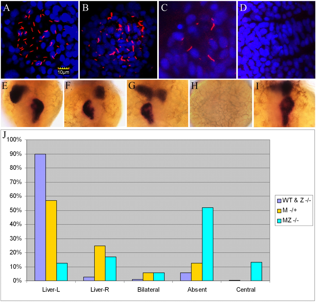
Fig. 7 Disruption of left-right asymmetry in Mta3 and MZta3 mutants. (A-D) Confocal images of embryos stained with anti-acetylated tubulin (red) and DAPI (blue) showing Kupffer’s vesicle in wild-type (A) Mta3 (B,C) and MZta3 (D). Note the variable number and length of motile cilia in Mta3 embryos compared with wild type. (E-I) Examples of liver and exocrine pancreas orientation revealed by in situ hybridization for the liver marker l-fabp and the pancreas marker trypsin. Orientations are categorized into five phenotypes: left liver and right pancreas, typical of wild-type (E), right liver and left pancreas (F), bilateral (G), absent (H), and centralized liver and pancreas (I). (J) Frequency distribution of different phenotypes among wild-type and ta3/ta3 embryos, and Mta3 and MZta3 mutant embryos.