
|
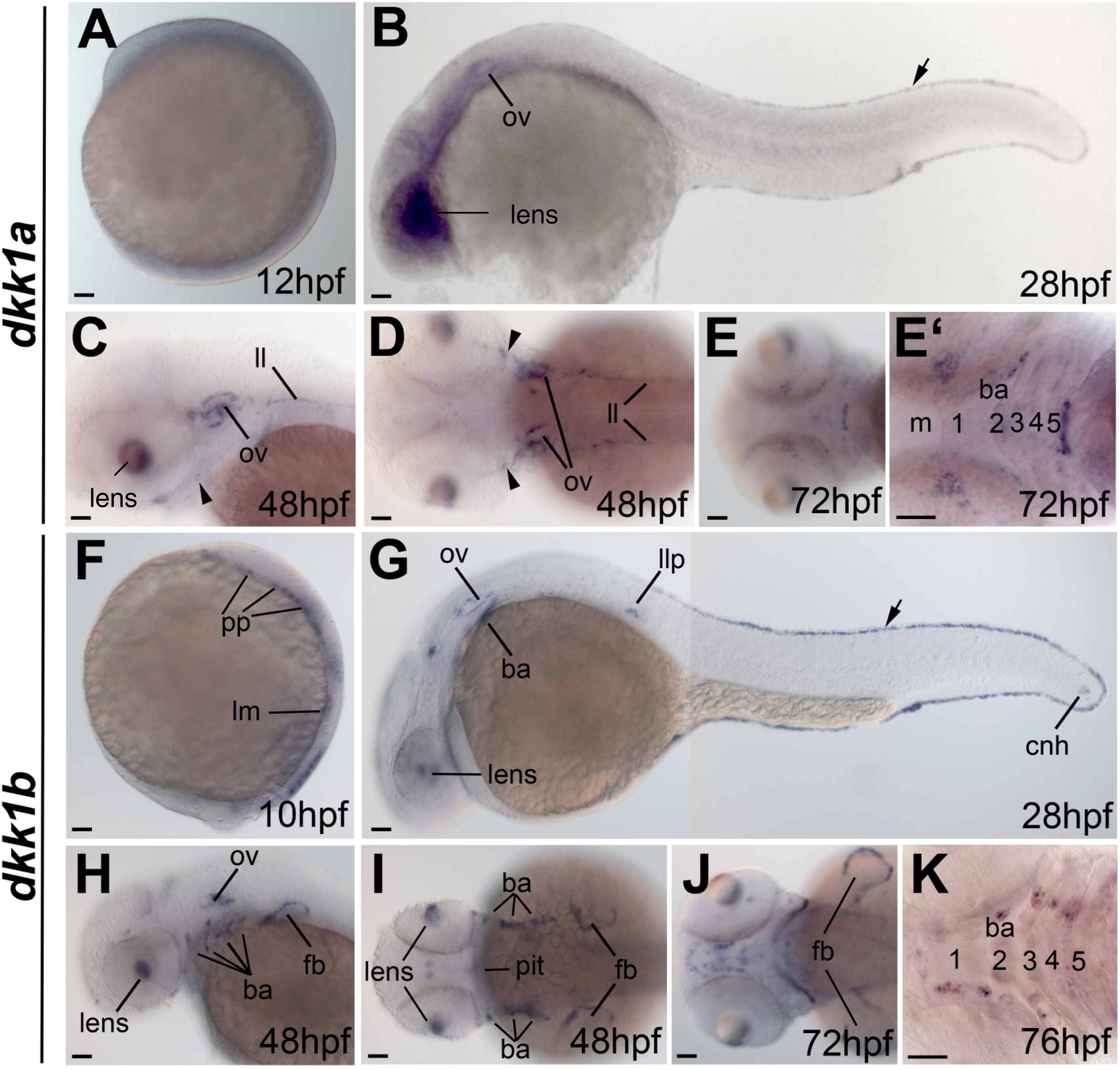
Fig. 2
Expression pattern of dkk1a and dkk1b during embryonic development Whole mount in situ hybridization analyses with dkk1a (A–E2) and dkk1b (F–K) specific antisense probes in developmental stages ranging from 10 to 76hpf. E2 and K show branchial arches of embryos in higher magnification. Arrowheads in C and D indicate position of individual dkk1a-expressing cells. Note the similar pattern of dkk1a and dkk1b expression in the epidermal edge lining of trunk and tail (arrow) and in the otic vesicles (ov) and lens. Distinct expression patterns of dkk1a were found in lateral line (ll) and of dkk1b in fin buds (fb), prechordal plate (pp), lateral posterior mesoderm (lm), chordaneural hinge (cnh), pituitary (pit), lateral line primordium (llp) and in the late embryonic branchial arches (ba; numbers indicate position the pharyngeal arches 1–5). Embryos are shown in lateral (A–C and F–H) or dorsal view with anterior to the left (D, E, E2, I, J and K). Scale bars in all figures correspond to 50 μm.
Reprinted from Gene expression patterns : GEP, 11(8), Untergasser, G., Martowicz, A., Hermann, M., Töchterle, S., and Meyer, D., Distinct expression patterns of dickkopf genes during late embryonic development of Danio rerio, 491-500, Copyright (2011) with permission from Elsevier. Full text @ Gene Expr. Patterns