
|
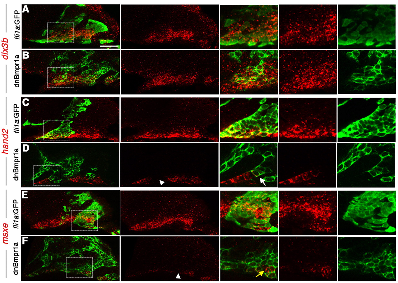
Fig. 4 Cell-autonomous regulation of DV gene expression by BMP. (A-F) Confocal sections of anti-GFP staining (green) and dlx3b (A,B), hand2 (C,D) and msxe (E,F) expression (red). Merged and individual channels are shown, as well as higher magnification views of boxed regions. Wild-type hosts received CNCC precursor transplants from either wild-type fli1a:GFP (A,C,E) or hsp70I:dnBmpr1a-GFP (B,D,F) donors. hand2 and msxe were cell-autonomously reduced in hsp70I:dnBmpr1a-GFP clones (white arrowheads), whereas dlx3b was largely unaffected. In high magnification views, the white arrow indicates a hsp70I:dnBmpr1a-GFP clone displaying loss of hand2, and the yellow arrow indicates a small clone of wild-type host cells still expressing msxe. Scale bar: 50 μm.