
|
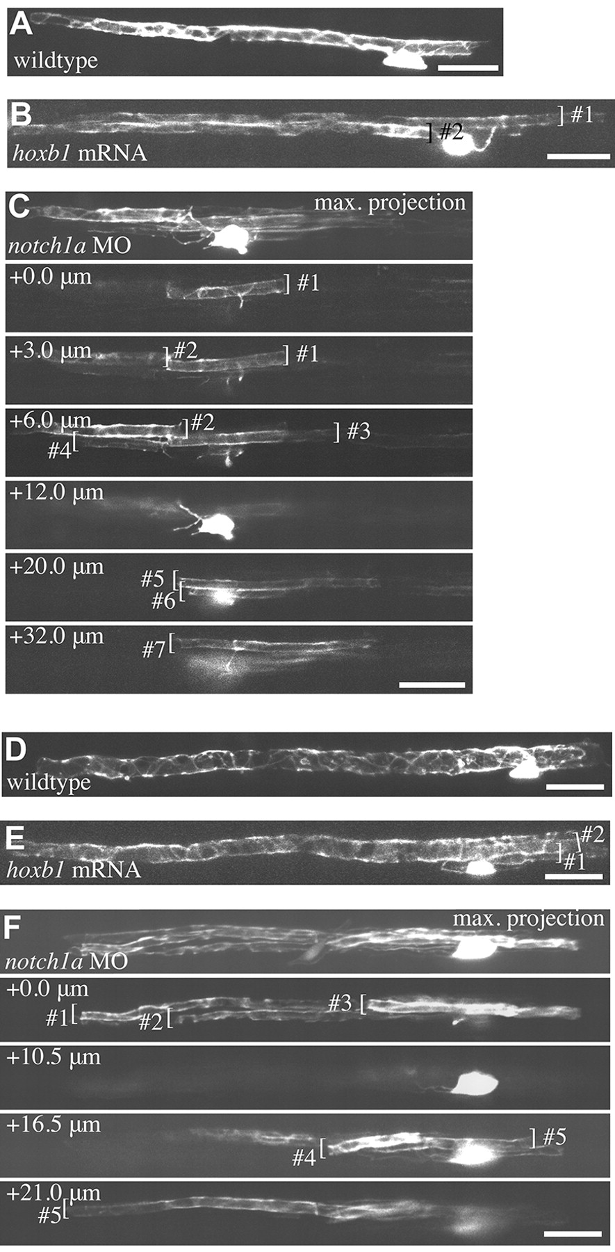
Fig. 6 Individual oligodendrocytes myelinate multiple supernumerary Mauthner axons. (A-F) All images are of live single mbp:EGFP-expressing oligodendrocytes in the spinal cord of larvae at 5 dpf (A-C) and 9 dpf (D-F). (A,D) Wild-type oligodendrocytes associated with single Mauthner axons. (B,E) Oligodendrocytes in hoxb1 mRNA-injected animals with myelin sheaths on two Mauthner axons. (C,F) Oligodendrocytes in notch1a morphants that make seven (C) and five (F) myelin sheaths on supernumerary Mauthner axons. Maximum intensity projections and individual confocal z-sections are indicated for clarity. Individual myelin sheaths indicated by brackets. Scale bars: 20 μm.