
Image
|
Figure Caption
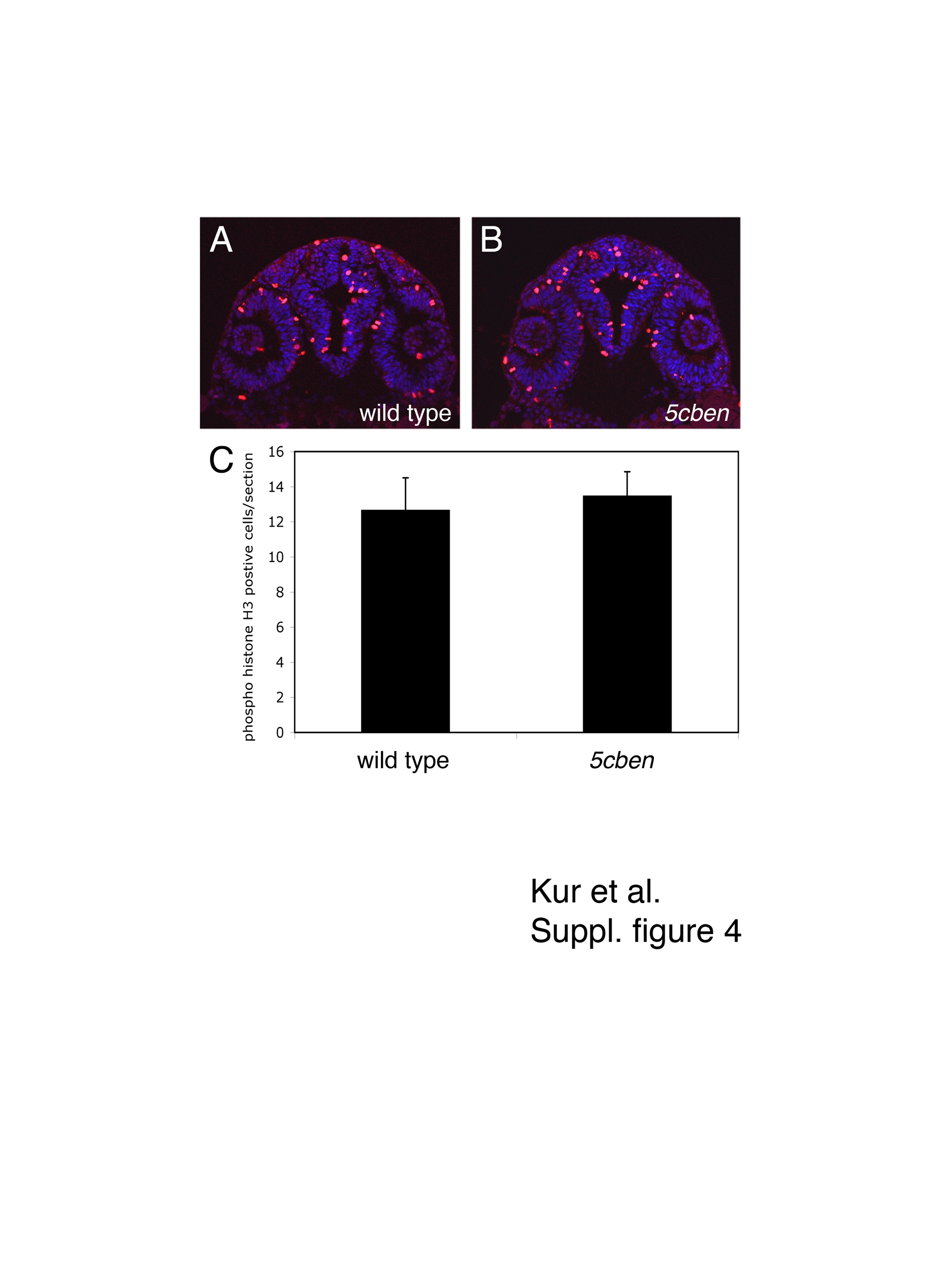
Fig. S4
Quantification of proliferation in the developing zebrafish forebrain.
(A, B) Immunohistological detection of phospho-histone H3 (pH3) in the diencephalon of wild type and 5cben embryos at 24 hpf. (C) Quantification of the number of pH3 positive cells per section (± SEM). n=3-6 sections each of a total of 10 embryos per genotype.
Acknowledgments
This image is the copyrighted work of the attributed author or publisher, and
ZFIN has permission only to display this image to its users.
Additional permissions should be obtained from the applicable author or publisher of the image.
Full text @ Dev. Dyn.