
|
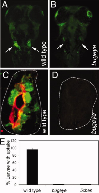
Fig. 4
Lrp2 deficiency disrupts clearance pathways in the pronephric duct. A, B: Whole mount fluorescence microscopy detects accumulation of fluid phase marker FITC-dextran (green) in the wild type but not in the Lrp2-deficient pronephros (arrows in A and B) upon injection into the common cardinal vein. C, D: Immunohistological detection of Lrp2 (red) and FITC-dextran (green) in the pronephros of the indicated genotypes. White lines highlight the position of the pronephric ducts. E: Wild type as well as bugeye and 5cben larvae at 4 dpf were injected with FITC-dextran and the number of larvae with tubular accumulation of tracers evaluated by fluorescence microscopy. Data are given as % ± SEM of all larvae injected. N= 53 (wild type), 15 (bugeye), or 23 (5cben).