
|
Fig. S4
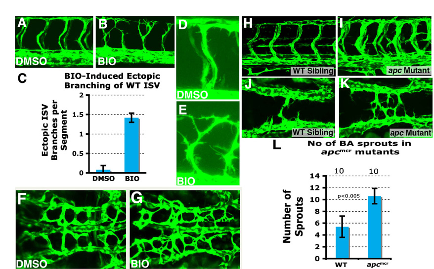
Ectopic vascular branching in BIO treated wild-type and Rspo1 morphant embryos. (A,B) Confocal images of the trunks of 40 hpf wild-type Tg(fli:EGFP)y1 zebrafish embryos treated from 16 hpf to 40 hpf with either carrier DMSO (A) or BIO (B). BIO-treated animals show enhanced ISV branching compared with their control DMSO-treated siblings. (C) Quantification of the number of ectopic branches per segment in trunks of 40 hpf wild-type Tg(fli:EGFP)y1 zebrafish embryos treated from 16 hpf to 40 hpf with either carrier DMSO or BIO. (D,E) Higher magnification confocal images of the trunks of 40 hpf wild-type Tg(fli:EGFP)y1 zebrafish embryos treated from 16 hpf to 40 hpf with either carrier DMSO (D) or BIO (E), showing increased protrusive activity in BIO-treated embryos compared with DMSO-treated controls. (F,G) Confocal images of the central arteries of 48 hpf Tg(fli:EGFP)y1 wild-type zebrafish embryos treated with DMSO (F) or BIO (G). (H,I) Confocal images of the trunk vessels of 48 hpf wild-type sibling (H) and homozygous apcmcr (I) mutant embryos. (J,K) Confocal images of the hindbrain cranial vessels of 30 hpf wild-type sibling (J) and homozygous apcmcr (K) mutant embryos. (L) Quantitation of number of basilar artery sprouts at 30 hpf in wild-type sibling and homozygous apcmcr mutant embryos.