
Fig. 5
|
Fig. 5
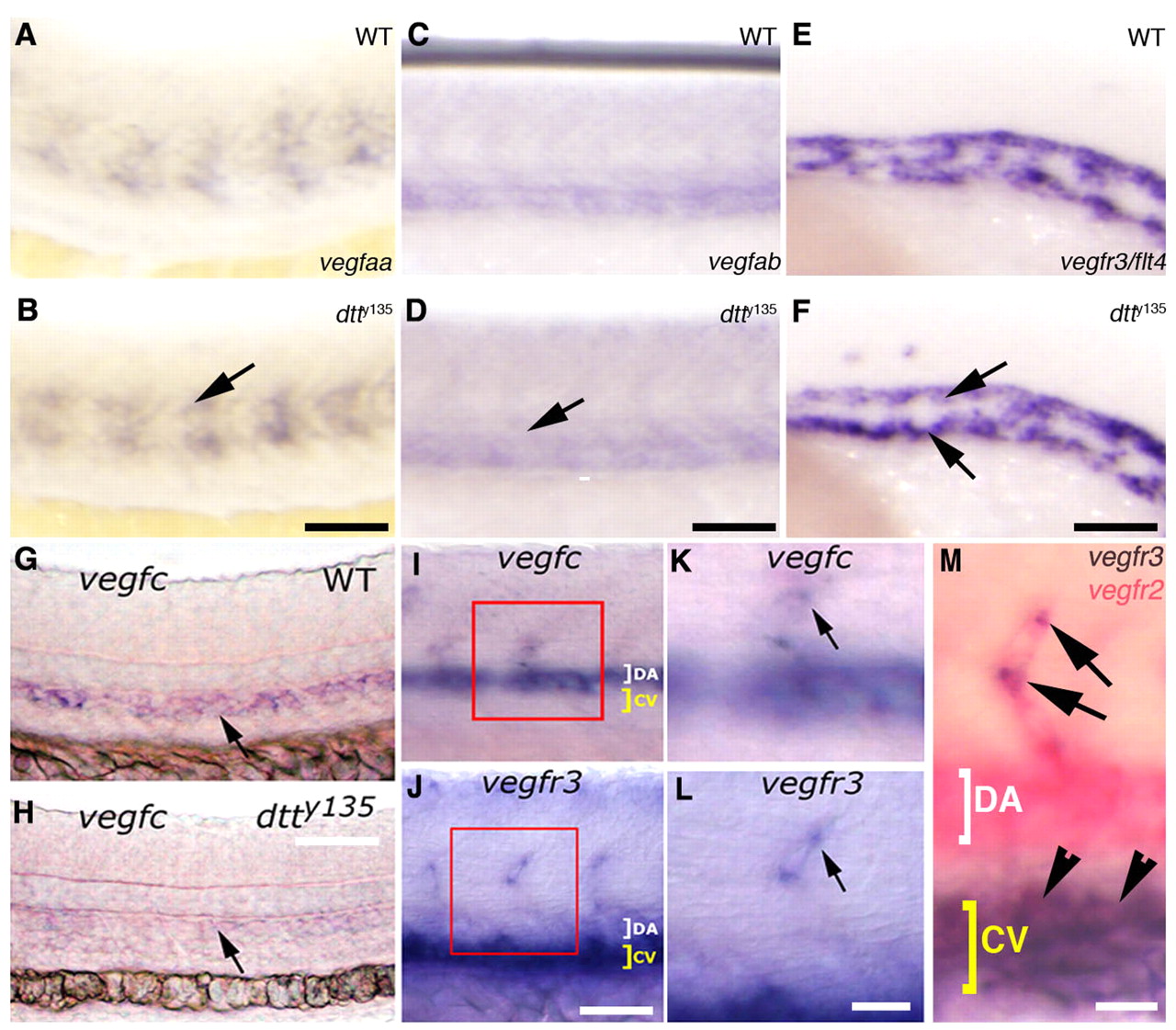
Expression of Vegfc is specifically affected in dtty135 mutant embryos. (A-F) In situ hybridization of the mid-trunk of wild-type sibling (A,C,E) and dtty135 mutant (B,D,F) zebrafish embryos probed for vegfaa (A,B), vegfab (C,D) or vegfr3 (E,F). The expression of vegfaa in the somites (arrow in B), vegfab in the axial vessels (arrow in D) and vegfr3 in the axial vessels (arrows in F) does not change in dtty135 mutant embryos compared with their wild-type siblings. (A-D) 24 hpf; (E,F) 20 hpf. (G,H) In situ hybridization of 26 hpf wild-type sibling (G) and dtty135 mutant (H) embryos probed for vegfc (arrows). (I,K) In situ hybridization of 26 hpf wild-type embryos probed for vegfc, showing expression in the DA and ISV (K shows a magnified image of the boxed region in I). (J,L) In situ hybridization of 26 hpf wild-type embryos probed for vegfr3, showing expression in the CV and ISV tip cells (L shows a magnified image of the boxed region shown in J). (M) Double in situ hybridization confirming expression of Vegfr3/flt4 (purple) in the tip cell (arrows) in addition to the cardinal vein (arrowheads), when compared with expression of Vegfr2/flk1 (pink) throughout the DA, CV and ISV. Scale bars: 50 μm in B,D,F,G; 45 μm in I,J; 25 μm in K-M.