
|
Fig. 5
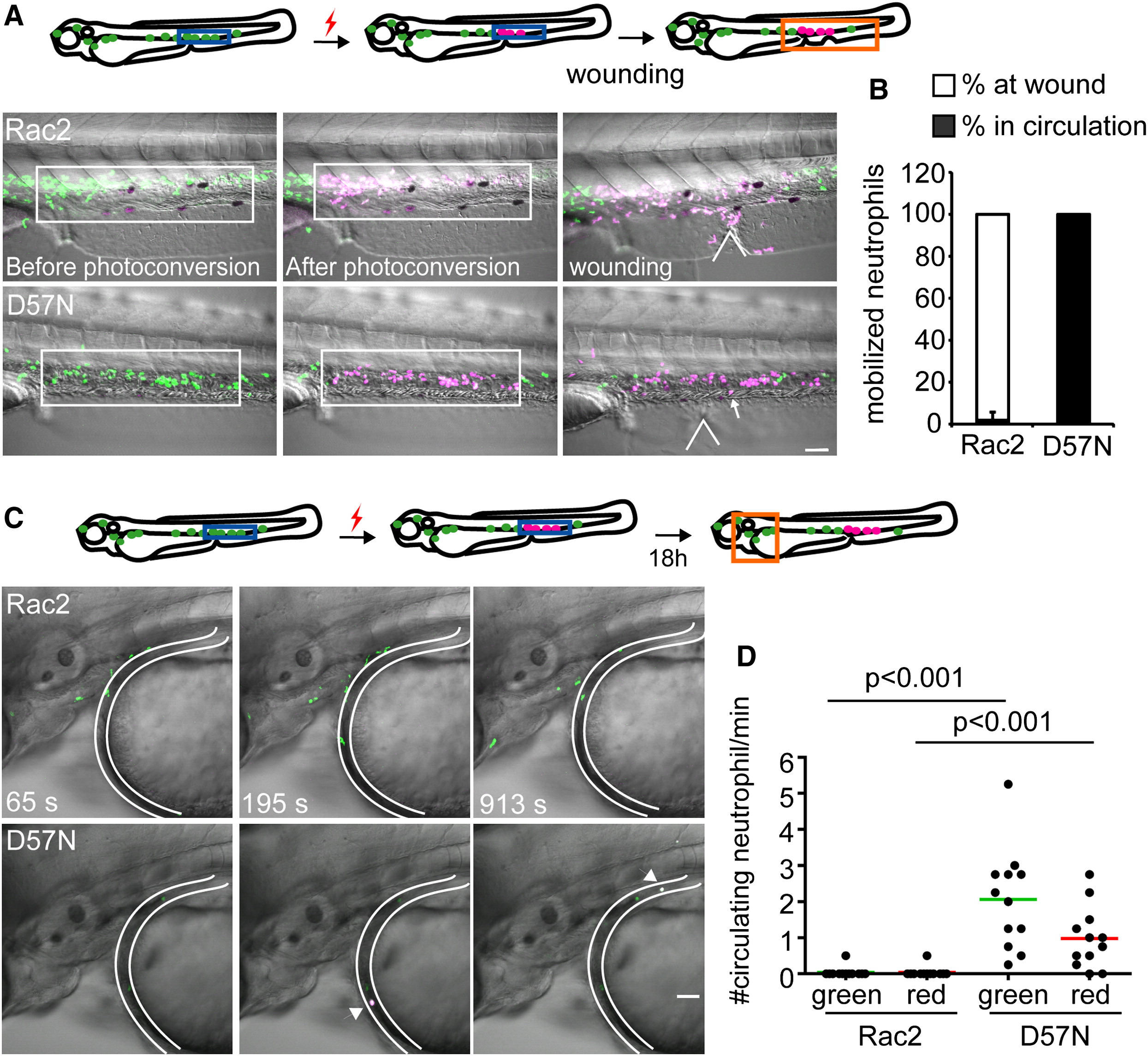
Rac2D57N Results in Increased Release of Neutrophils from the CHT
(A) Rac2 or D57N lines were crossed with Tg(mpx:dendra2). Photoactivation converts green fluorescent Dendra2 to red, allowing for fate-mapping 1.5 hr postventral fin wounding. Neutrophils in boxed region (CHT) were photoconverted. Locations of wounds were indicated with the wedged lines. Arrow, a circulating neutrophil. Scale bar represents 50 μm.
(B) Distribution of photolabeled neutrophils that are mobilized out of the CHT in Rac2 and D57N larvae 2 hpw. Open bar, % of photolabeled neutrophils at wound; filled bar, % of photolabeled neutrophils in circulation. n = 6 larvae for each condition, results are presented with mean ± SD.
(C) Neutrophils from the CHT in 2 dpf D57N larvae spontaneously entered the circulation 18 hr after photolabeling. Still images from representative movies in the head are shown. Location of blood vessel is delineated. Arrow, circulating photoconverted neutrophils. Scale bar represents 50 μm.
(D) Quantification of (C). Green and red represent total and photolabeled circulating neutrophils, respectively. n = 12 each. p < 0.001, Kruskal-Wallis test followed by Dunn′s multiple comparison test.
See also Figure S4 and Movie S7.
Reprinted from Developmental Cell, 21(4), Deng, Q., Yoo, S.K., Cavnar, P.J., Green, J.M., and Huttenlocher, A., Dual roles for Rac2 in neutrophil motility and active retention in zebrafish hematopoietic tissue, 735-745, Copyright (2011) with permission from Elsevier. Full text @ Dev. Cell