
|
Fig. 4
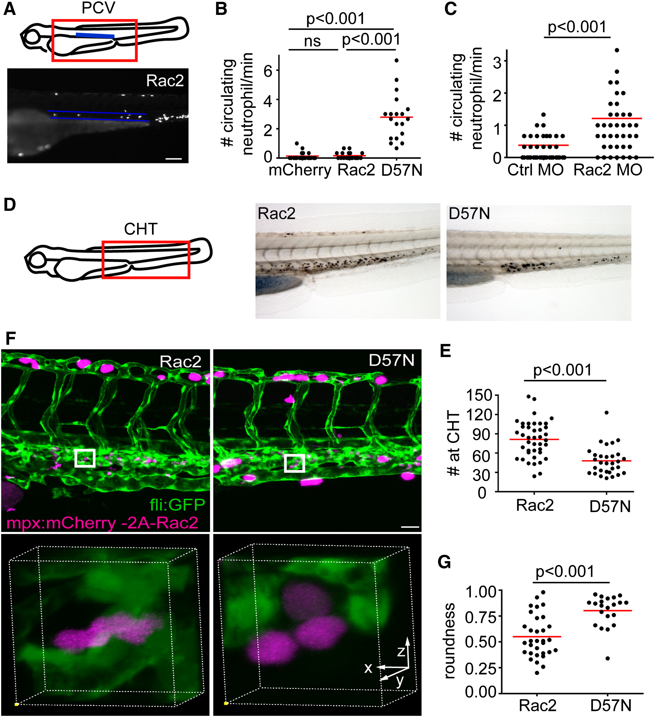
Neutrophilia in Rac2-Deficient Larvae
(A) Illustration of the location of the posterior cardinal vein (PCV) that was imaged for quantification of circulating neutrophils. Neutrophils that circulate through the PCV per minute were scored. Lower, one still image from the representative movie of Rac2 larvae containing circulating neutrophils within the highlighted PCV (blue lines). Scale bar represents 100 μm.
(B) Quantification of neutrophils that circulate through the PCV per min in mCherry, Rac2, and D57N larvae at 3 dpf. n = 20 each for mCherry, Rac2, and D57N larvae. p < 0.001, Kruskal-Wallis test followed by Dunn′s multiple comparison test.
(C) Quantification of neutrophils that circulate through the PCV per min in control or Rac2 morphants at 2 dpf. n = 40 each for Ctrl or Rac2 morphants. p < 0.001, two-tailed Mann-Whitney U test.
(D) Sudan black staining of neutrophils in the CHT.
(E) Quantification of neutrophil number in the CHT of Rac2 and D57N larvae at 3 dpf. n = 41 (Rac2) and 30 (D57N). p < 0.001, two-tailed Mann-Whitney U test.
(F) Three-dimensional volume rendering of neutrophil morphology in the CHT in Rac2 and D57N larvae. Boxed regions are enlarged in the lower panel. Founder fish are crossed with Tg(fli1:GFP) to visualize endothelial tissues or stroma. Scale bar represents 30 μm.
(G) Quantification of cell roundness in the CHT in Rac2 and D57N larvae. n = 32 (Rac2) and 23 (D57N). p < 0.001, two-tailed Mann-Whitney U test.
See also Figure S3 and Movie S6.
Reprinted from Developmental Cell, 21(4), Deng, Q., Yoo, S.K., Cavnar, P.J., Green, J.M., and Huttenlocher, A., Dual roles for Rac2 in neutrophil motility and active retention in zebrafish hematopoietic tissue, 735-745, Copyright (2011) with permission from Elsevier. Full text @ Dev. Cell