
|
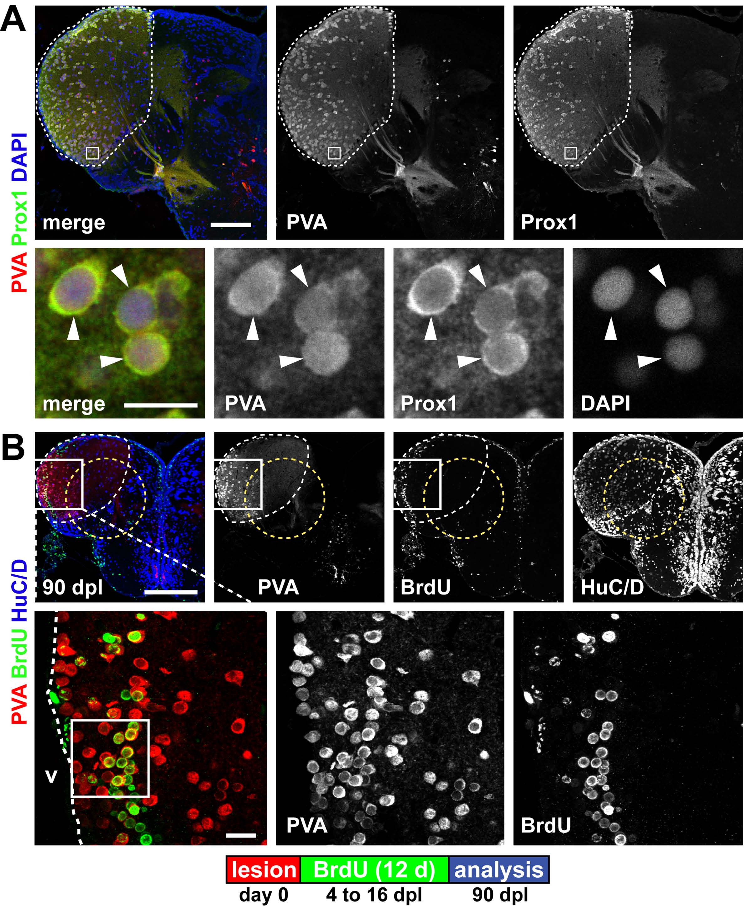
Fig. S9 Parvalbumin and Prox1 expression mark a territory within the lesion site in the dorsolateral telencephalon. (A) A territory in the dorsolateral telencephalon is characterized by the co-expression of the interneuron marker parvalbumin (PVA, red) and Prox1 (green) in a neuronal subpopulation (white dashed outline). Co-expression (arrowheads) is shown in a single confocal section. (B) The PVA/Prox1 territory (white dashed outline) is further characterized by weak expression of HuC/D (blue) and reaches into the lesion canal (yellow dashed circle). When BrdU is applied 4 to 16 dpl to label newborn cells, many periventricular PVA+ (red)/BrdU+ (green) are found at 90 dpl within the dorsolateral telencephalon, as shown in confocal max projections. This shows that the lesion does not alter the fate of newborn neurons: they acquire a spatially appropriate neuronal subtype. The area framed in the inset indicates the location of the single confocal section displayed in Fig. 5A. Scale bars: 200 μm; 50 μm in inset.