
|
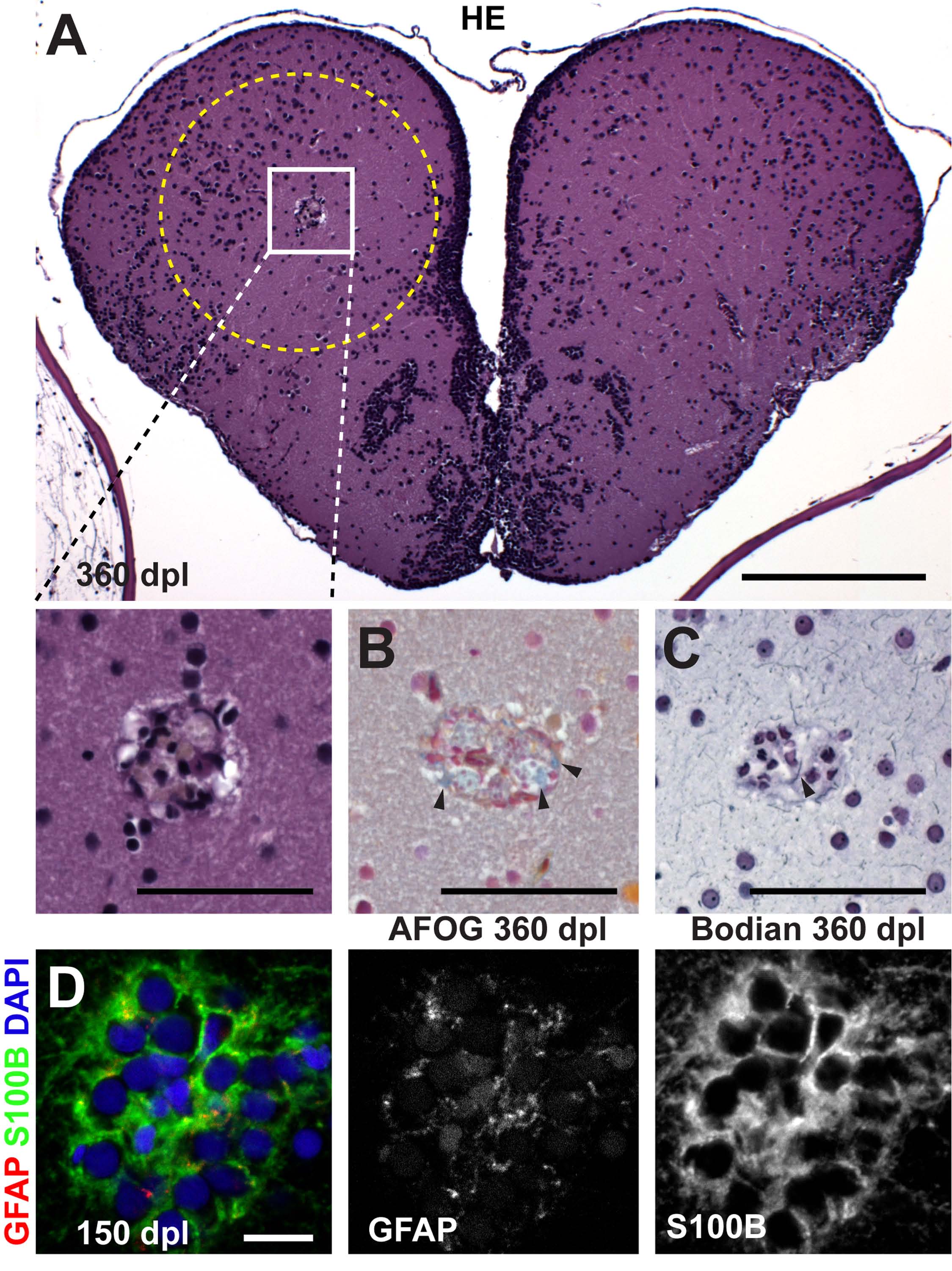
Fig. S6 A small confined glial inclusion is remaining in stab lesioned hemispheres after long survival times. (A) At 360 dpl in two out of three fish a 30×40×30 μm3 cyst is seen in the caudal dorsal telencephalon in HE-stained 1 µm paraffin sections that may be a remnant of the previously much larger lesion canal that has otherwise closed. (B) Acid-Fuchsin/OrangeG staining (AFOG) shows extracellular matrix (arrowheads, collagen,blue) deposition within the remnant 360 dpl. (C) Neuronal processes can be detected around and within (arrowhead) the glial inclusion by Bodian silver staining 360 dpl. (D) At 150 dpl, the inclusion consists of cells that are weakly positive for GFAP (red) and strongly express S100B (green), identifying their glial/ependymal character (nuclei, DAI, blue), as shown in single confocal sections. Scale bars: 200 μm in A, 50 μm in inset; 50 μm in B,C; 10 μm in D. The dashed outline represents the lesion canal.