
|
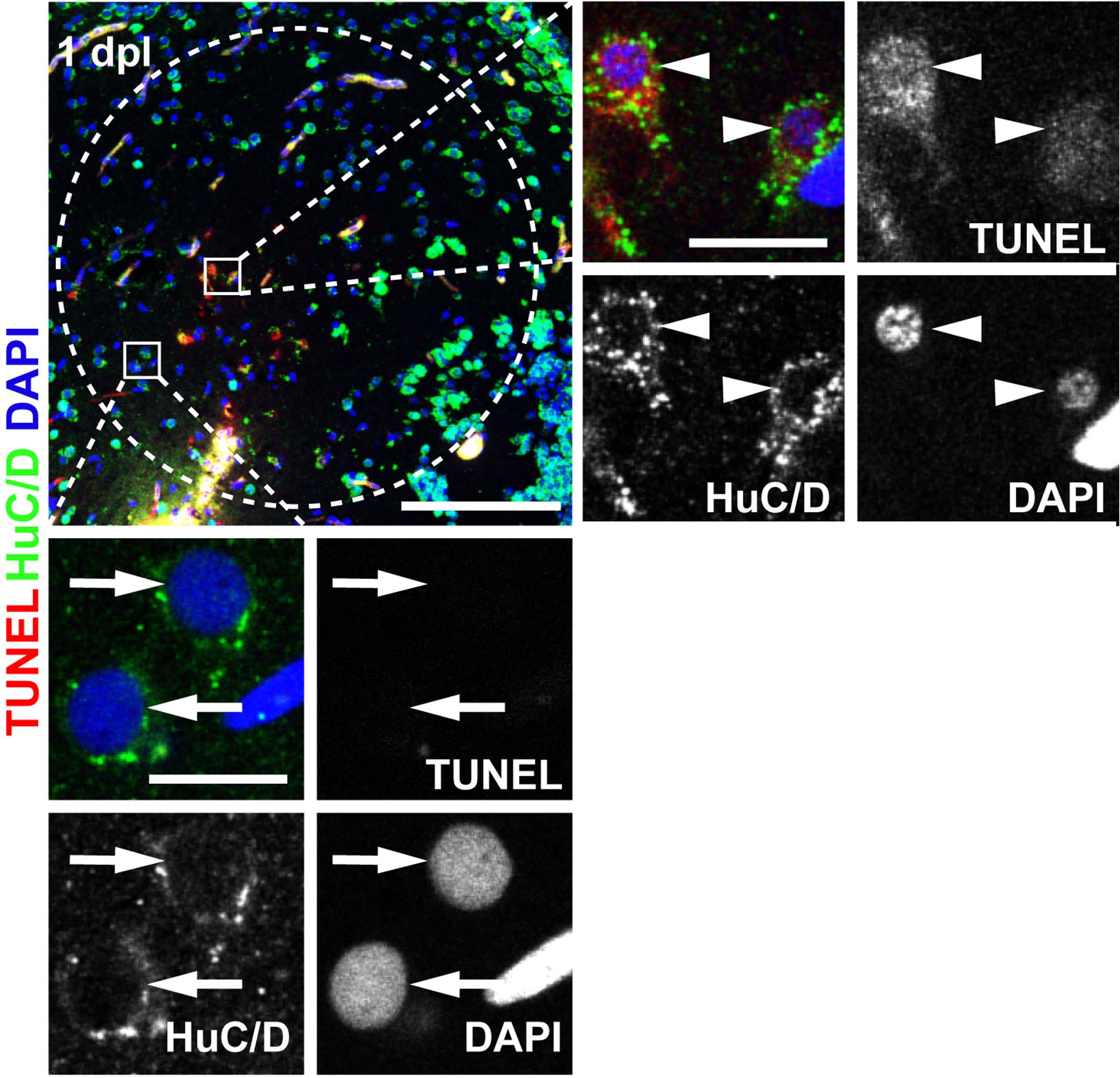
Fig. S3 Neurons within the lesioned hemisphere enter cell death rapidly after lesion. TUNEL+ (red)/HuC/D+ (green) double-positive, dying neurons (arrowheads) are found in the lesioned parenchyma (right inset panel) at 1 dpl. Most dying cells show hallmarks of apoptosis, like cytoplasmic condensation and condensed pycnotic nuclei (DAPI, blue, compare dimensions with TUNEL-, not dying neurons, shown in the inset panel below) and also characteristics of necrosis, such as nuclear and cytoplasmic TUNEL signal. Note also the spotty and weak HuC/D-signal that is a hallmark of damaged neurons. Insets show single confocal sections. Scale bars: 100 μm and 10 μm in insets respectively. Dashed outline represents the lesion canal.