
|
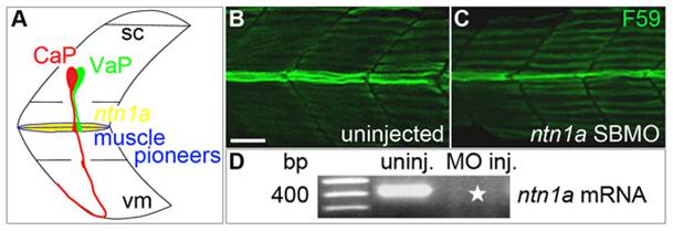
Fig. 1 Ntn1a expressed in the muscle pioneers is a candidate signal for stopping VaP axon outgrowth.
A. Schematized view of one spinal hemisegment including spinal cord (sc) and overlying muscle. CaP (red) and VaP (green) axons contact muscle pioneers (blue) at the first intermediate target. The VaP axon stalls at the muscle pioneers while the CaP axon continues into ventral myotome (vm). ntn1a mRNA (yellow) is expressed in muscle pioneers [16]. This image and following images are oriented laterally with rostral to the left. B–C. Projected confocal stacks of (B) uninjected and (C) ntn1a SBMO-injected embryos labeled with F59, a slow muscle fiber marker [70]. At 24 hpf, muscle pioneers (green fibers in the middle of the images) are present in both control and MO-injected embryos. D. RT-PCR results confirming uninjected embryos have wildtype ntn1a (450 bp band) that is absent from embryos injected with 5.6 ng of ntn1a SBMO (star). Multiple samples were run in this gel. Lanes containing samples unrelated to ntn1a were cropped from the figure. Scale bar = 20 μm.